Puzzles in the Pattern of Plague

This past weekend, my husband and I attended a lecture at my university on ‘Puzzles in the Pattern of Plague’. Being an infectious diseases specialist in my day job, and having talked about plague a number of times on the blog, the topic really caught my attention. As my husband is also a science geek, he good naturedly tagged along.

The talk was about using mathematical modeling of historic plagues to be able to predict future epidemics. And before you starting thinking Ugh, math… it was a very down to earth talk without a smidge of calculus (though you know for a fact that this kind of modelling overflows with calculus in the background), so it was fascinating from both a historical and scientific viewpoint. We’ll come back to a modern standpoint at the end as we touch on SARS and what the outbreak in 2003 might have meant for humanity.

Dr. David Earn, a mathematician at McMaster University, was the evening’s speaker. He focused on plague—bubonic, septicemic, and pneumonic—with an emphasis on the Great Plague of London from 1665.

There have been multiple waves of plague over the past two millennia:

  • Justinian, starting in 541CE and lasting 200 years.
  • Black Death (which includes the Great Plague of London), originating in China in 1334 and lasting more than 350 years, finally ending in the late 1600’s after killing over 60% of Europeans.
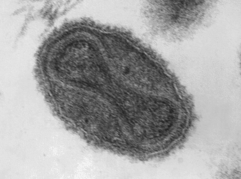
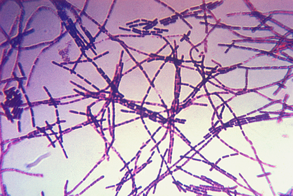
  • Modern plague, which started in China in 1860 before spreading to kill 2–10 million worldwide, but which finally enabled scientists of the day to isolate the responsible bacterial agent, Yersinia pestis (Y. pestis).

There are three main forms of plague known to modern man:

  • Bubonic plague—an infection of the lymphatic system by Y. pestis leading to swollen lymph nodes, or buboes. If untreated (as it was historically), the death rate is approximately 66%. Treated with modern antibiotics, the death rate is approximately 11%.
  • Pneumonic plague—a Y. pestis infection that spreads to the lungs resulting in pneumonia. Untreated, the death rate approximates 100%. Surprisingly, even with modern antibiotics, the death rate is still nearly 100%. This is the kind of plague that defense experts worry about as a bioterrorism threat. There is a vaccine, but it is extremely inefficient.
  • Septicemic plague—a Y. pestis infection which enters and spreads via the circulatory system leading to blackened and gangrenous extremities. The death rate from septicemic plague falls between that of bubonic and pneumonic plagues, but tends to approximate that of pneumonic plague even with modern treatment.

When it comes to identifying the type of plague through the ages, historians have no choice but to fall back on records of the time, which are often vague, and mostly date after the Justinian plague. However, it is clear that at least part of the second wave of plague was likely partially septicemic since it was known at the time as the Black Death, a reference to the black fingers and toes resulting from the systemic infection.

Considering the differences in plague properties, how can we be sure that the Justinian, Black Death, and modern plagues were caused by the same modern Y. pestis agent? Another McMaster researcher, Dr. Hendrik Poinar, has sequenced ancient DNA found in plague victims from both of these epidemics and has confirmed that Y. pestis was responsible for both.

So how does mathematical modelling help us use plagues of the past to possibly save us during a plague of the future? Information is power, and, in this case, knowledge of previous pandemics can help us design better control strategies for the next pandemic. This paradigm could instruct future scientists and healthcare professionals how to interrupt an epidemic just as it starts, possibly saving millions of lives in the process.

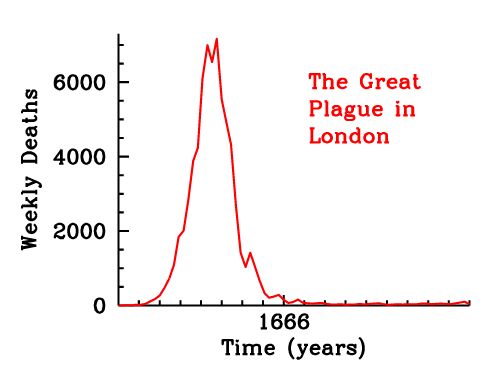
Toward this end, Dr. Earn and his students examined the Great Plague of London of 1665. They went back to the weekly bills of mortality published during that time period to collect large scale data. This is a written record of not only all the deaths broken down by cause, but also where within the 130 parishes the deaths occurred, and exactly how many were caused by plague. Based on this data, they were able to show in great detail how the plague ripped through London during the summer of 1665 and then simmered for the next year before finally disappearing at the end of 1666 following the Great Fire of London. The plague was mostly gone by the time of the fire, but actually continued for several more months, so the fire didn’t contribute to its disappearance.

London weekly mortality register, September 12 – 17, 1665 (click for a larger version)

Mathematicians use the susceptible/infectious/removed (SIR) model to infer transmission and recovery rates during an epidemic. In the SIR model

  • S = susceptible, the number of people who could be infected.
  • I = infectious, those who are infected and are capable of passing on the pathogen.
  • R = removed, those who have either recovered and are now immune, or who have died. In either case, these are the people who are now taken out of the susceptible population.

Using this SIR model, R0, the reproduction number (how contagious a pathogen is) is calculated. R0 essentially describes how many secondary cases can arise from a single primary disease case. If one sick person can spread disease to only one other person, then R0 = 1. If, on average, the disease spreads to 2 people, then R0 = 2 etc. For disease to spread through a population, R0 must be greater than zero, or there is not enough transmission to maintain the epidemic. To put this in perspective, influenza R0 = 1.5–3 and measles R0 = 17 (so get your kids vaccinated, parents! With an R0 like that, herd vaccination will only take you so far…).

Dr. Earn was able to calculate the difference between Black Death plague as it spread from Asia and through Europe in the following years: 1348, 1361, 1375, 1563, 1593, 1603, 1625, and 1665. What they found was that the R0 for plague actually rose over the centuries with an R0 = 1.1 in 1348 and an R0 = 1.5 in 1665. That translated to only 20% infection in the 14th century, but 50% infection in 1665 by the numbers. But they had to make one major assumption for this calculation: that the transmission rate in the second plague pandemic was similar to modern plague and the modern bacteria that scientists have studied. And knowing the historic death rates, Dr. Earn knew that assumption had to be incorrect since over 60% of the population was infected.

So what could account for this difference? And why did the plague spread nearly twice as fast in the 16th century, compared to the 14th century? We’ll never know for sure, but several possibilities exist:

  • The pathogen itself may have changed and become stronger through mutations that were later lost when they no longer conveyed a survival advantage.
  • Population density likely played a role as people started to live in dense clusters with close contact inside city walls.
  • Climate change of the time also played a role, as the Little Ice Age occurred between 1300–1850 in Europe.
  • What form of plague dominated at the time since bubonic had a much higher survival rate than either the septicemic or pneumonic variants.

So how does this information serve us in modern times? Dr. Earn cited the SARS pandemic of 2003 as an example. In Canada, we had 250 infections, from which 50 patients died, so a 20% mortality rate (which is quite severe in modern times with modern drugs). Worldwide, over 8,000 people were infected with 774 eventual deaths. However this pandemic could have been much worse but for a number of factors. China, the original location of the outbreak, reacted very quickly, and used extreme isolation of anyone diagnosed with SARS or who had come into contact with it. It spread through air travel to limited locations, with Canada being the next worst hit. I remember the SARS pandemic very clearly as our lab was located in the university hospital and coming to work every day included extremely long lines, written questionnaires, one-on-one health screening, and a single monitored entrance through the parking garage. But it was procedures like patient isolation and absolute dedication to stopping spread that halted the pandemic in its tracks and no infection has been seen since 2004. However, Dr. Earn calculated the SARS R0 = 2, which would have led to a 50% infection rate. In his estimation, had we not been able to control the outbreak, it would have quickly gone worldwide with over one billion dead. Simply stated, it would have changed the course of human history just as the 14th centuryplague changed the course of history after killing 50 million, just over half the population.

So this research is critically important. Mathematical models show that even if there had been a plague vaccine during the Great Plague of London that only protected 5% of the population, it would have made a significant change to the transmission curve and millions would have been saved. Using these tools, mathematicians will be able to assist during pathogen outbreaks as healthcare professionals are making decisions around treatment and vaccination and how to best protect the population and save the most lives. I’ve said for years that the bugs are going to win someday, but tools like this could stave off that fate.

Interested in more of what Dr. Earn and his team do? He gave a TEDx talk a few years ago, and this clip shows some of the fascinating animations he showed us last night about the spread of the Great Plague through the burrows of London:
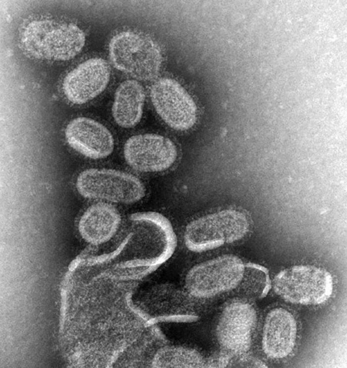
Canine DNA Profiling

Ann and I are back to blogging now, but we're also shifting back into more forensics-related posts as we're moving toward the release of LAMENT THE COMMON BONES, book five in the Abbott and Lowell Forensic Mysteries. Today, we're looking at a topic that spans our two series as we examine the forensic technique which recently saved the life of a service K-9.

DNA profiling has been used in law enforcement, medical examiners, and archeologists for humans for decades. DNA is used for profiling both victims and suspects in crimes, for identifying the dead after mass disasters, and tracing family lineages through mitochondrial DNA. But the same techniques can be used for other species.

Recently a case came to light of a Belgian Malinois service dog named Jeb who was sentence to be destroyed after he was convicted of killing a neighbour’s dog, Vlad. He was not actually witnessed killing the 16-pound Pomeranian, but he was found by the late dog’s owner standing over the body of the dead dog. While not definitive, it certainly didn’t look good for Jeb. He was taken into custody by Animal Control and a judge was appointed to hear the case. After hearing testimony, including how the neighbour was scared by the large dog because ‘he always barked’, the judge made the reluctant decision to designate Jeb as a ‘dangerous animal’. As a result, he had no choice but to call for the dog’s death.

However the owners, Penny and Kenneth Job, never believed for a moment that their dog was capable of such a violent act. They had adopted Jeb after he’d been rescued as an abandoned pup in Detroit by their daughter, Kandie Morrison. Morrison worked for a local rescue group, but quickly recognized that the young Malinois would make an excellent service dog for her father, as United State Air Force veteran with Charcot-Marie-Tooth, a neurodegenerative disease. With the help of a local veterinarian, Jeb was trained into a gentle, dependable service dog to help support Ken Job and to be there for him if he falls.

The accusation of Jeb being the cause of Vlad’s death didn’t make sense to the Jobs. This simply wasn’t the dog they knew and who lived with three dogs and seven cats in complete peace. Rather than simply taking the heartbreaking news at face value, they took matters into their own hands. While they had previously believed Vlad had been cremated following the investigation, they discovered during the course of the trial that his body was instead frozen. They had argued during the trial that a stray dog had been seen in the area around the time of the killing, and the area was populated with wild foxes, but now they had a chance to scientifically prove Jeb’s innocence. They swabbed the inside of his cheek and arranged for samples to be taken from Vlad’s wounds for comparison at the Maples Center for Forensic Medicine at the University of Florida College of Medicine.

The Jobs were thrilled when the results came back vindicating their dog. Yes, Vlad has been killed by a dog, but not by Jeb. Shortly after, Jeb was released and returned to his loving family and crucially important life of service. DNA had proven his innocence, exonerating him just as it can exonerate innocent human convicts.

DNA analysis is not a regular part of canine cases, even those that call for the destruction of an animal. But the $460 spent by the Jobs definitely decided the case and saved the life of their beloved pet and helpmate. Perhaps it’s time to start thinking about animal cases in the same light—when a life hangs in the balance, isn’t it worth ensuring that justice is being done? $460 doesn’t seem like that high a price to pay to avoid an innocent paying for a crime he didn’t commit.

Photo credit: CNN

The Bugs Are Going To Win… Or not?

First of all, our apologies for being AWOL for the last several weeks. We were on deadline on the developmental edit of LONE WOLF and barely had time to sleep, let alone come up with a well-researched article for the blog. But the manuscript is done now, and handed in, and we’re back in business!


Normal
0


false
false
false

EN-US
X-NONE
X-NONE


 <w:LatentStyles DefLockedState="false" DefUnhideWhenUsed="true"
DefSemiHidden="true" DefQFormat="false" DefPriority="99"
LatentStyleCount="267…

A T4 bacteriophage poised to infect a bacterium.

I have a saying that I seem to repeat often: “The bugs are going to win.” This refers to the oncoming crisis humankind is likely to have against microbes (or ‘bugs’ as we generically refer to bacteria in the lab). In a day and age when factory-farmed animals are given antibiotics prophylactically and too many medical professionals still consider knee-jerk prescription writing for antibiotics an acceptable act, news stories repeatedly warn of the number of strains of bacteria resistant to all antibiotics. Another continual theme are reports of now-resistant bacteria that used to be susceptible to a shrinking number of heavy hitting antibiotics used in only the most severe hospital cases.

In many ways, the thought of these tiny organisms taking out human kind is hard to wrap your brain around. We’re the top of the food chain; we have technology and science on our side. How can they top that? Part of the answer is very simple—replication rate. Your average bacteria divides into two identical daughter cells approximately every 20 minutes. It takes humans approximately 20 years for that same replication. This rate of bacterial replication leads to a very large number of progeny—in one day, a single bacterium can produce 4.7 x 10^21 bacteria. Even if the mutation rate is only 1 in 10^10 DNA base pairs, depending on the genome size of the bacteria, hundreds of mutations can occur in a span of less than 10 hours. Some of these mutations could lead to an inactive bacterium, but one of them might lead to a new gene conferring resistance to one of our last ditch antibiotics.

What happens when we can no longer control bacteria with a pill or injection and we enter the post-antibiotic age? It will be like going back to the early 20th century, before the advent of penicillin. Pneumonia would be a fatal infection. Bacterial epidemics similar to the Black plague that killed 30 million people—a full third of Europe at the time—could once again be a scenario faced by the world. Those who are very young and very old will be at risk due to compromised or immature immune systems. It’s a very scary concept.

So how can we beat the bugs?

  • Develop new chemical therapies or multi-therapies: Scientists at Merck Research Laboratories in New Jersey recently announced two separate compounds, tarocin A and tarocin B, that makes Methicillin-resistant Staphylococcus aureaus (MRSA; a bacteria well known for its resistance) susceptible to antibiotics again. Tarocins A and B target the bacterial cell wall, and, when used with standard antibiotics, kill the bacteria effectively in mice. Human trials will be the next test.
  • Find new natural anti-bacterial compounds: Dr. Gerry Wright at McMaster University has been sending students around North America to sample soil in different areas. Compounds from those soils are then tested against bacteria. If the bacteria are killed, the samples are then further tested to determine the successful compound. So far the team has made two notable discoveries:
    •  AMA, a molecule produced by a fungus in Nova Scotia soil that is able to knock out one of the strongest and most worrisome antibiotic resistance genes, NDM-1. NDM-1 is such a concern that the World Health Organization identifies it as a global health threat. When AMA is used, regular antibiotics once again become effective.
    • Teixobactin is an antimicrobial molecule produced by one bacteria to kill others and was discovered in the soil from a field in Maine. The compound has been shown to be effective in killing multiple antibiotic resistant strains of bacteria.
  • Revisit OLD remedies: A one thousand year-old Anglo Saxon recipe to treat eye infections using onion, garlic, wine and cow bile has been found by modern scientists to kill MRSA. Rather than being any individual ingredient that was effective, it was the combined recipe that killed 90% of the MRSA in the sample.
  • Use their own natural enemies against them: We’re all familiar with the concept of viruses. Bacteriophage are viruses that attack bacteria (see the above photo), acting as all viruses do—they inject their contents into the bacterium and highjack it’s replication machinery to produce hundreds or thousands of copies of itself, eventually bursting the bacterium, killing it. Scientists in Russia and parts of Europe have used bacteriophage for nearly a century as antibacterials. The Western world is now looking at phage therapy as a potential replacement for antibiotics.

In the end, the question remains can we beat the bugs, and that remains to be seen. In the meantime, be conscientious citizens of the world. Do not demand antibiotics for your kids because they have ear infections, of which a full 70% can be caused by viruses; wait and let the infection run its course and it will likely clear up just as fast without antibiotic usage. Don’t run to the doctor every time you have a cold until you hit the 7 to 10 day range and a secondary infection may have set in. We should only use antibiotics when they are truly necessary. And maybe, just maybe, the bugs won’t win after all.

Photo credit: Xvivo Scientific Animation

Zika: Emergence of a New Epidemic

Electron micrograph of Zika virus particles.

Electron micrograph of Zika virus particles.

You’d have to be living under a rock for the last few weeks to have missed the fact that a new virus—called Zika—has been making headlines. In reality, Zika is not a new virus at all, however its effects in the last six months in Brazil and other south American countries has been utterly alarming. Worst, one of its most terrible of its outcomes strikes the most fragile of us—unborn infants.

The Zika virus was discovered in 1947 in the Zika Forest in Uganda. It is one of a number of viruses in the Flaviviridae family which includes dengue virus and West Nile virus. By far, the main method of transmission is via the mosquito Aedes aegypti, therefore the areas of mosquito-borne Zika infection follow the natural distribution of Aedes aegypti in an equatorial band around the planet. Historically, disease caused by Zika has been considered mild and mostly trivial with four out of five infected people never knowing they were ever infected. Those who develop symptoms usually present with a low grade fever, joint and muscle pain, and headaches lasting from approximately two to seven days. Since there is no effective medicine or vaccine, symptoms are treated directly—rest, plenty of fluids, and acetominophen to ease the aches and pains. For most patients that is sufficient, and severe disease and death were extremely rare.

However, a disturbing picture emerged from Brazil last November when the news broke that 4,000 babies had been born in 2015 with microcephaly—a birth defect in newborns with abnormally small heads and incomplete brain development. Compared to the normal average of 150 babies born with microcephaly in a single year, this is a staggering increase. While we’re still waiting for definitive evidence that this increase is due to the Zika virus, Zika was first identified in the country in May of 2015, and some pregnant women at the time showed evidence of the virus’ RNA genome, leading public health officials to infer that Zika was responsible. Late last week, Columbia reported three people who died of Guillain-Barré syndrome—a disorder where the body’s immune system attacks the peripheral nerves causing muscle weakness and paralysis—following Zika infection.

Recently, the World Health Organization took the significant step of defining Zika as a ‘Public Health Emergency of International Concern’. However, it is difficult to make recommendations about Zika since the virus is not well studied. Less than 200 scientific papers mention Zika, only a handful of which are notable for significant information. And the fact that so much is still unknown—all methods of transmission, life cycle of the virus, where it replicates inside the body, and what body fluids it can be found in and for how long—ties the hands of public health officials who are trying their best to prepare people on how best to protect themselves. The virus has been found in saliva, blood, semen, and urine, leading to warnings about sexual transmission, pregnant woman kissing anyone other than their partner, and to blood banks setting restrictions on blood donations in a window following travel to Zika-ravaged countries. Some countries, like El Salvador, have even suggested to women that they refrain from getting pregnant for the next two years, a difficult issue in a Catholic country where most methods of birth control are forbidden. At this point, the main preventative strategy is to avoid travel in the twenty-six countries with documented Zika infections. If not, avoidance of mosquito bites is the next best thing, by using bug spray, wearing long sleeves and pants to cover the skin, and sleeping in rooms with window screens or air-conditioning.

This story has struck a personal note in my day job as a manager of an infectious diseases lab. Flaviviruses are one of our main areas of study, with past projects including major NIH studies of both West Nile and dengue fever. In fact, dengue virus is carried by the same mosquito as Zika, opening up the possibility of co-infection or super infection (infection of a second virus following infection of the first). While the West Nile study was of North American cases, the more recent dengue fever study involved cases and controls from Central and South America and southeast Asia. Currently, we’re looking at the tens of thousands of samples in our freezers from that study and wondering how many of them might also contain Zika. Discussions are already well under way and we hope to join the fight against Zika very soon. We’re certainly better situated to hit the ground running than most labs considering our existing sample bank, and we have high hopes that we can make a meaningful contribution.

So where do we go from here? First and foremost, remember all the public health agencies are doing their best to keep people safe. It’s a damned if you do and damned if you don’t situation—people will be upset if recommended precautions seem too severe, or if they turn out to be too lax as additional information is discovered.  They’re doing their very best based on extremely limited information. And while for the majority of people, Zika is not likely to be a major health concern, for the sake of those that are more significantly affected, it behooves us all to be diligent to protect the vulnerable.

Photo credit: Wikimedia Commons

Amazing Genetic Tales: Chimeras

A chimera mouse with two of her non-chimeric offpsringA story hit the news last week that was meant to be an interesting human vignette, but the forensic aspects of it immediately jumped out at me. Ah, crime writers. Sometimes we see the world through a special lens!

The story begins with an American couple who conceived a child through in vitro fertilization. The mother carried to term and gave birth to a healthy baby boy. Unexpectedly, however, the boy’s blood type didn’t match either parent, and they became concerned a mix-up had occurred at the fertility clinic. The clinic maintained that on the day of the donation, the father was the only white man to donate sperm; since the child was clearly white, no mix-up had occurred. Still the couple wanted full tests run, so the father contributed a saliva sample and a paternity test was run which concluded that the boy was not his son. Needless to say, the parents were devastated, but they requested a more detailed test through the commercial genetic ancestry company 23andMe.

No one anticipated the results of that test. It was revealed that the man was not the boy’s father, but was instead his uncle. As we’ve discussed in the past, standard identification by DNA is established using 15 markers, but 23andMe uses a genotyping method (near and dear to my heart as we’ve just finished a 5-year Dengue study in the lab based on this technique) called GWAS—Genome-Wide-Association Study—to look at hundreds of thousands of genes for the purpose of building a detailed ancestry map. Because of this extremely thorough analysis, they were able to determine that the boy was the nephew of the man thought to be his father.

However, the man didn’t have a brother. So there was only one conclusion to be drawn from the analysis. Keep in mind the statistic that 1 in 8 single births start as multiple pregnancies, but one of the children is lost very early and, rather than being miscarried, is simply reabsorbed in the womb. Sometimes these cells are then incorporated into the surviving child, making that child a chimera—an organism made up of cells originating from genetically distinct individuals. The man must have been the only survivor of what were originally two fraternal twins, as absorbing an identical twin would have been indistinct from his own natural genotype. As a result, the sperm he produced carried his unborn brother’s genetic signature, but his saliva carried his own. It’s the first known case in the world of a chimera fooling a paternity test.

As a biologist currently heavily involved in complex genetics and genotyping, I was instantly interested in the details of this case. But as a crime writer, I immediately considered the forensic implications of this gentleman. Not that I’m suggesting he’s going to take this information and suddenly adopt of life of crime, but any man with this type of chimerism could be a rapist that would be beyond normal law enforcement’s ability to apprehend since typical DNA sampling techniques would not capture his true genetic state. Luckily, this is a very rare genetic occurrence, and though TV crime shows like CSI might use this scenario as a plot device, the chances of it happening in real life are exceedingly small.

Photo credit: Wikimedia Commons

Rewriting the History of the Black Plague

We’ve discussed the history and science behind the Black Death several times already on Skeleton Keys—from bubonic plague victims discovered in London, to a cemetery under a Parisian supermarket, to whether rats were truly responsible for spreading the epidemic, to the 15th century Bedlam hospital cemetery that was recently unearthed, and finally to sequencing Yersinia pestis, the bug that caused the disease. But the plague was back in the news again last week with the announcement of the discovery of Bronze Age skeletons containing DNA from Y. pestis, indicating the existence of the plague a full 3,000 years earlier than originally suspected.

While the most well-known instance of plague is the 14th century Black Death epidemic that ravaged Europe and killed more than half the population (approximately 50 million people), the earliest known epidemic was in 6th century Germany. And while theories hold that the ancient Greeks also experienced plague, there is no scientific proof of its existence in that population.

The Bronze Age is known as being a time of not only the development of much stronger bronze tools by smelting copper with tin and other metals, but also the development of early writing systems and the first structured (though early) civilizations. It is also known as the time of a sudden mass migration from Russia and modern day Ukraine into Europe. Scientists now think they can explain why.

DNA extracted from the teeth of 101 Bronze Age skeletons was sequenced in hopes of finding traces of Y. pestis as a method of explaining the mass exodus. To their surprise, a significant number of specimens (7%, which is high as a single cause of illness-based death in a normal general population) contained Y. pestis sequences, and two specimens contained sufficient DNA to encode the entire Y. pestis genome. The oldest strains dated back to 3,000 B.C., a full three millennia before previously theorized plague origins.

When scientists studied the stains of Bronze Age Y. pestis and compared it to the deadly 14th century version, they found an interesting evolutionary tale. The earliest versions of the plague bacteria lacked the gene that enables the bacteria to colonize the gut of fleas which enables them to be the vector between human hosts. Without that insect vector, Y. pestis could only be spread human-to-human directly through blood or saliva, and, as such, was much less transmissible. However, by 1,000 B.C., that gene was present in the bacteria allowing for zoonotic (animal/insect to human) transfer and increased rates of infection. These same early Bronze Age versions of the plague contained another gene, one that allowed the bacteria to infect the lungs of humans. As a result, Bronze Age Y. pestis likely caused pneumonic plague rather than bubonic plague (an infection of the lymphatic system). This was by no means a preferable version of the disease—while infections were less common, the death rate from pneumonic plague was 90 - 100%, as opposed to bubonic plague’s 30 – 90%.

Such a catastrophic death rate supports the theory that a mass migration occurred in an effort to escape the ravages of the disease. Without the advantage of transmission and transport via fleas—which would use other animals to move from place to place, often in the company of potential human hosts—Bronze Age people could successfully escape the disease by traveling into Europe. DNA studies of Europeans have previously confirmed a shift in genetic makeup from typical European hunter gatherers in 3,000 B.C. toward the Yamnaya phenotype typical of the Russian/Ukrainian area, around 2,000 B.C.

Photo credit: Nature

Biosecurity Incidents In Top U.S. Labs—What, Me Worry? Smallpox Edition

Smallpox is one of the few diseases that modern medical science has managed to eradicate. Believed to have emerged around 10,000 B.C., the disease first appeared in historical documents in the 15th century. The disease is estimated to have killed 300 to 500 million people in the 20th century alone, having a fatality rate of 20–60% in adults and 80% in children.

Smallpox is widely taught in university immunology classes as the world’s first vaccine. Edward Jenner noticed in 1798 that milkmaids who developed cowpox seemed to be immune to smallpox infection. In a stunningly unethical, off-the-cuff experiment, when a milkmaid came to him for treatment, he ran a thread through one of her pustules, coating it with pus. He then inoculated the eight year old son of his gardener by making a small cut and running the pus-coated thread through it. Shortly after, the boy became symptomatic for a mild case of cowpox. Several months later, Jenner took pox scabs from someone with small pox and similarly inoculated the boy a second time. The boy was immune to smallpox and remained healthy. This was the beginning of the vaccine revolution.

In 1967, the WHO mandated the eradication of smallpox, using newer, modern vaccines based on vaccinia virus, a virus related to both smallpox and cowpox. The last known case of smallpox occurred in 1977, and the WHO considered it eradicated in 1979.

This left the world with only laboratory strains of the virus. Following a breach of containment, resulting in the death of a lab worker in 1978, any labs with remaining virus either destroyed them or transferred them to safer labs. Currently, only the U.S. Centers for Disease Control and the Russian State Research Center for Virology and Biotechnology still retain samples. The argument has been made that all remaining virus should be destroyed, but the existing aliquots are retained in case any other stocks arise leading to a dangerous bioweapon in the wrong hands. At this point, decades after the final vaccinations, essentially everyone except military personnel (who continue to be inoculated with the vaccinia vaccine for out of country work) would be susceptible to a fresh onslaught of smallpox. But with only two stocks of the virus in the world, we’d like to believe that we’re safe.

So it was somewhat of a shock in July 2014, when the National Institutes of Health reported that six glass vials of freeze-dried smallpox stock had been found in a long forgotten box in the back of a cold storage room. I remember hearing the news and being stunned for several reasons—glass vials to store a biosafety level IV pathogen (of course, there was no sterile, disposable Nalgene polypropylene cryovials back then, but glass? So incredibly dangerous…), no security, and no inventory so no one even knew they were there. The stock was estimated to have been there since the 1950s, even though the building didn’t open until the 1960s, and from the 1970s on was used by the Food and Drug Administration. A further investigation reveals twelve boxes in total containing smallpox, dengue, influenza, Q fever, and rickettsia, all previously unknown to be stored there, and all with no security precautions (proper security precautions would involve a minimum of two locked doors between the pathogen and the public, detailed inventories, and full biosafety training of all personnel). The FDA immediately mandated a full review of all cold storage spaces to ensure no other pathogens were present.

The glass vials were immediately transferred to the CDC to undergo testing, where it was determined that two of the six vials contained viable virus. Had the tubes broken, the world could have seen a smallpox pandemic the likes of which it hadn’t seen for decades and for which we are entirely unprepared. Luckily for all of us, the vials remained intact. All the vials were destroyed following testing.


Ann and I are going to be taking some time off for summer holidays and to really concentrate on drafting LONE WOLF, book one of the new FBI K-9 Mysteries with Kensington Books. So we look forward to coming back fresh and with a lot of solid writing behind us in September. See you then!

Photo credit: Wikimedia Commons

Biosecurity Incidents In Top U.S. Labs—What, Me Worry? Influenza Edition

In the same report published by the CDC in July 2014 that discussed a laboratory incident concerning anthrax, an incident concerning influenza was also revealed. In August 2014, a full report detailed the cross-contamination of a non-pathogenic H9N2 strain of avian (bird) influenza with the highly pathogenic H1N1 strain (remember the 2009 flu pandemic? That’s the one…) which then sent it out to a U.S. Department of Agriculture lab that had no idea what they were dealing with. Fortunately, the Department of Agriculture lab is also a biosafety level III (BSL III) lab, so the samples were handled under BSL III containment procedures. As it turned out, no workers were infected with the pathogenic strain, a very lucky break as the story could have ended very differently.

As a scientist, the moment I heard this story, I knew exactly what had likely happened and it’s a big no-no in working with cells and viruses. Keep in mind, the error wasn’t identified until six months after the fact, so the worker couldn’t recall the day like it was the previous week, but reported working with then non-pathogenic H9N2 virus, decontaminating the biological cabinet and the working with the pathogenic H1N1 virus. However, of the 1.5 hours required to do all of this, key card access indicated the worker was only present for 51 minutes and that also included time to shower out of the facility and dress in street clothes. Clearly, the full protocol was not carried out. The scientists admitted that they were under pressure at the time to complete work for an upcoming WHO vaccine conference, and, that day in particular, the scientist in question was rushing to get to a lab meeting. Corners were clearly cut.

There are two scenarios that could have happened:

  1. Considering that each infection should take 30 minutes, it is possible the scientist did both infections in the biological cabinet at the same time. To put it plainly, this is NOT done. One of the first rules of tissue culture is that products are kept separate to ensure purity of the product and safety of the current and any future lab worker. I’d like to think this isn’t what happened.
  2. Instead of following protocol and working with the less pathogenic strain first and then the pathogenic strain, the scientist may have mixed up the order, working with the more pathogenic strain first and then not decontaminating afterward before moving onto the second, less virulent strain. Personally, I think this is what happened. Also, as PCR testing of the H9N2 strain doesn’t indicated H1N1 contamination, it’s likely they weren’t used concurrently.

One other concerning incident happened in association with this cross-contamination. When the receiving lab started to use the virus, chickens in the experiment started to unexpectedly die. Upon testing their virus, they determined that their H9N2 was contaminated with the deadly H1N1. When they informed the CDC, the lab team tested their stock of H9N2 to confirm that it was indeed contaminated with H1N1. But they did not report the incident at the time. It wasn’t until a second CDC team found atypical results with their stock of H9N2 virus that the original team reported the incident. At that time, all connected stocks of H9N2 were destroyed. Luckily all work done by the second team was conducted under BSL III containment, so there was no risk to any of the lab personnel.

For a group that is considered by most scientists as the gold standard, this incident combined with the anthrax incident is quite distressing. Scientists did not display good laboratory practices, training, communication skills, and in many cases, common sense. As a scientist myself, especially as one trained in BSL III procedures, many of these errors would simply not be acceptable or expected at our facility. Protocols and training are already in place to avoid this and a detailed incident reporting procedure is in place (and let me assure you, I’ve used it for BSL III incidents twice). After all this, I know I certainly look at the CDC differently, and I’m sure many other scientists do as well.

Next week we’ll be back with our last installment in this series. What happens when a virus that is nearly eradicated pops up in someone’s freezer? Come back next week and we’ll tell you all about it…

Photo credit: Wikimedia Commons

Biosecurity Incidents In Top U.S. Labs—What, Me Worry? Anthrax Edition

Today we’re continuing on with our series on laboratory biosafety and how it’s gone wrong lately in places you would think would be immune to such incidents. But it just goes to show human error can trump every precaution you put in place and that hopefully your people would follow to the letter. Especially when lives are on the line. But not so much, apparently…

In July of 2014, the Centers for Disease Control (CDC) held a press conference to discuss two different incidents that had taken place the previous month. We’re going to showcase one incident this week and one next week.

The first incident involved a CDC lab preparing extracts from anthrax, a biosafety level III (BSL III) pathogen. Their usual protocol involved chemically deactivating the pathogen for 24 hours, but they learned of a new protocol that only required a 10 minute deactivation. Now, this protocol was not for anthrax, but was instead for Brucella, another BSL III pathogen. However, they elected to try this new protocol on anthrax. This method also included a double check—after the pathogen is deactivated, some of it is plated onto agar plates and incubated for 48 hours to ensure there is no growth, and all the pathogen is dead. The CDC first attempted this protocol in June of 2014, sampling the extract to agar plates after 10 minutes, but leaving the rest for the full 24 hours.

However, due to a misunderstanding during a phone conversation and a lack of follow-up with the actual printed protocol, the scientist responsible only left the agar plates for 24 hours post deactivation, at which time, the plates were determined to have no growth. The scientist intended to autoclave the plates and discard them that day, however he could not open the autoclave door, so the plates were returned to the incubator. At this time, the anthrax incubated for 24 hours was distributed as deactivated pathogen to biosafety level II (BSL II) labs. Eight days later, the agar plates were removed from the incubator for disposal and, to their surprise, anthrax growth was observed. Scientists realized the 10 minute procedure was not sufficient to kill anthrax, but were unsure if the 24 hour procedure (from which anthrax had been sent out to BSL II labs and their lower level of containment) was sufficient. At that point, it had to be considered that they had a breach in biosafety containment and all the labs had to be completely decontaminated. Later tests showed that the 24 hour procedure was sufficient to kill most of the anthrax, but not all. As a result, while it was unlikely that infection would occur, it was not impossible.

Upon closer examination, there were a number of issues that led up to this incident:

  1. Use of unapproved techniques—there were several related to filtering of the extract, but this also included observing the plates for sterility at 24 hours instead of the required 48 hours.
  2. Transfer of material not confirmed to be inactive—based on the error made in point 1, this also involved a lack of written protocol of what was required to ensure that pathogens were truly deactivated.
  3. Use of pathogenic strains of anthrax to test out a new protocol when non-pathogenic strains would have revealed the same conclusion with none of the risk. This was an extremely unwise decision.
  4. Inadequate knowledge of peer-reviewed literature by both the laboratory scientist and his supervisor—papers already existed outlining that this method was not sufficient for absolute sterility of infectious anthrax.
  5. Lack of standard operating procedures to document pathogen deactivation.

Fortunately, no staff member ever presented with symptoms of anthrax. But the laboratory in question was closed pending a number of assessments, the establishment of new procedures, and until remedial action was taken with the staff involved. This was not a shining moment for the CDC, the institution we scientists like to consider the gold standard in biosafety. This is the group you call when something goes wrong and this is what their own people are doing?

In the last month, a new anthrax story surfaced concerning an Army lab at the Dugway Proving Ground in Utah. It creates anthrax test kits and sends them out to numerous laboratories for local testing. Contained in the kit is a radiation-killed sample of anthrax to use as a positive control for testing. However, in May, a Maryland biotech company identified that live anthrax was present in the samples. Upon closer inspection, it appears the samples were not sufficiently treated to kill all the spores. The test kits were packaged and sent out, many by regular FedEx delivery, to 69 labs around the United States, but also in Canada, Britain, Australia and South Korea. The CDC has confirmed that all kits shipped between 2004 and 2015 contained live anthrax.

Even more disturbing information surfaced this past week. It appears that from 2007 onwards, the lab was aware that their deactivation process (chemical deactivation at the time, and later irradiation) was not sufficient to kill all the anthrax, but they ignored the issue. At the time, federal regulators at the Office of the Inspector General were aware and recommended further investigation and potential enforcement action, however nothing was ever done. Furthermore, the information was never reported to Congress—the group responsible for oversight of this lab—so they were completely unaware of the situation. No laboratory workers have shown symptoms of anthrax; however 31 workers are receiving antibiotics as a precaution.

Next week, we’ll be returning the CDC as we discuss a serious mishap concerning a highly pathogenic strain of influenza.

Photo credit: Wikimedia Commons

Biosecurity Incidents In Top U.S. Labs—Biosafety

This post may be a little off the beaten path for some of our blog readers, but it’s a story I’ve been watching over the past year or more. And when a new twist on the story hit the news in the last week, I thought it might be a good blog topic, even though it doesn’t have anything to do with writing or forensics. In this case, I’m looking at something near and dear to my day job—infectious diseases research.

In July, 2014, several alarming announcements came out of the U.S. Centers for Disease Control (CDC) involving potential and proven accidental exposures of lab personnel to extremely infectious pathogens. Then, that same month, the NIH announced the discovery of an even more dangerous pathogen found forgotten in FDA freezers. Just last month, the Pentagon admitted that one of its Army labs sent live pathogen out to as many as 69 different labs in a number of different countries. And now, this past week, it was revealed that same lab was secretly sanctioned for those actions eight years ago, but this information was hidden from Congress, which is responsible for oversight of the lab.

It’s enough to give a person nightmares about biological disasters.

Before we get into what went wrong with labs the world considers the gold standard in biosafety, let me give you a little bit about my background. I’ve worked at McMaster University in Hamilton, Ontario for nearly 25 years. I sat on the university’s Presidential Biosafety Committee for 5 years, making decisions on how to implement government standards for the safe use of pathogens, and how to rectify procedures that resulted in accident. I’ve worked nearly 25 years with biosafety level II pathogens (herpes simplex viruses I and II, vaccinia virus, Adenovirus, influenza, dengue virus and many others), and for over 15 years with biosafety level III pathogens (HIV and herpesvirus saimiri). Biosecurity has been my life for my entire adult working career.

What exactly does ‘biosafety level’ mean? The biosafety level of a pathogen is based on a number of factors: infectious dose (i.e. how much is required to contract the disease), mode of transmission (i.e. blood borne vs. air borne), host range (i.e. human vs. animal or both), the availability of effective treatment (i.e. pharmaceuticals), and the availability of preventative treatment (i.e. vaccines)

There are four internationally recognized biosafety levels for pathogens:

  • Biosafety level I—an organism unable or unlikely to cause disease in healthy individuals. May cause disease in immunocompromised individuals.
  • Biosafety level II—infectious organisms that cause disease, but are unlikely to make an individual seriously ill under normal circumstances. Effective treatment and preventive measures are available, and the risk of spread is limited. Example: herpes viruses, including those that cause mononucleosis, chicken pox and roseola.
  • Biosafety level III—infectious organisms that cause serious disease, but do not spread by casual contact. Organisms that cause diseases treatable by antimicrobial or antiparasitic agents. Example: HIV, anthrax.
  • Biosafety level IV pathogens—infectious organisms that cause very serious disease, often untreatable and leading to death, and are spread by casual contact. Example: Ebola virus.

Different containment levels are required to study pathogens at different levels.

  • Biosafety level I pathogens, like many strains of non-infectious E. coli, can be used on the open bench in the main lab.
  • Biosafety level II pathogens must be used within a separate level II room in a biological safety cabinet. Air is drawn into the cabinet away from the user and only exhausts into the room after HEPA filtration, thereby keeping any infectious particles trapped inside the cabinet. The user only interacts with the pathogen after donning a lab coat and gloves and by remote access i.e. using a pipettor to actually contact/transfer the pathogen.
  • Biosafety level III pathogens are used in an isolated, approved access-only laboratory with negative pressure similar to a biosafety cabinet—air is drawn in from outside the laboratory and only exhausts from a laboratory through giant HEPA filters in the ceiling. Workers inside the facility must wear rear-closing gowns and double gloves at all times. In the case of airborne pathogens, workers must also wear respirator hoods with a powered air purifier worn on the belt. A safety shower must be located in the laboratory in case of spills and pathogen appropriate disinfectants must be available at all times. In case of incident, detailed reporting and follow-up is mandatory.
  • Level IV pathogens can only be used in biosafety level III style labs situated in an isolated building. Work will involve all the requirements of biosafety level III work, but over and above that, the use of a positive pressure personnel suit (which pushes air out of the suit and away from the user) is mandatory at all times. Multiple showers (both in-suit and out), a vacuum room, and a UV room are required for clean exit from the facility.

Specialized training for each specific biosafety level is required by all personnel, often with annual updates and review. As you might imagine, the complexity of the training increases in orders of magnitude as the biosafety level increases.

Another aspect that is crucial to biosafety work, especially at upper levels, is tracking—where pathogen is stored, how it is treated, when small volumes are removed, how they are used, and how they are destroyed. There are strict guidelines concerning any transfers outside the laboratory as well—how it can be removed and who is allowed to receive it based on their facilities and training.

So now that you understand some of how this work is done, next week we’re doing to look at what went wrong (when it absolutely shouldn’t have) and what’s being done to safeguard workers and the public. See you then!

Photo credit: Wikimedia Commons

A Writer with a Day Job

Dengue_map.jpg

I’m going on a bit of a tangent this week and breaking away from my usual theme of forensics and writing to touch on another aspect of my life—my day job. Like many writers out there, writing may be my passion, but I have a responsibility to help my husband support our family financially, so I work full time. I worked in the field of HIV research for 20 years, but last winter my lab downsized, and I started looking for a new position. Last July, I joined a dynamic research group specializing in infectious diseases. We study a range of diseases, including pneumococcal infections and influenza, but our big project is a very large, international study of dengue fever.

In the map above, the areas in blue indicate the current risk for dengue virus infection, and the pins in red indicate areas where the disease is spreading or has been carried by travellers. Currently 40% of the world’s population lives in areas where the virus is endemic, leading to an estimated 50 million cases of dengue fever annually. Of those cases, 500,000 patients require hospitalization and 20,000 – 25,000 patients die of the disease. The virus spreads to humans by two types of mosquitos and the incidence of the disease matches the geography inhabited by these insects. Because of global warming and the northern spread of the Aedes Albopictus mosquito into the southeastern United States, the CDC has classified the dengue virus as a Biodefense Category A pathogen; a category that encompasses the most dangerous of the infectious diseases due to their easy transmission, high mortality rate and lack of effective treatment.

The large majority of patients infected with dengue virus show no symptoms at all or only present with a mild illness including fever, aches, a mild skin rash and joint pain—leading to the colloquial name for this disease: bone break fever. But approximately 5% progress to severe illness, and a subset of those exhibit life-threatening disease (dengue hemorrhagic fever and dengue shock syndrome), including symptoms such as platelet loss, abdominal bleeding, fluid accumulation in the chest, low blood pressure and organ dysfunction. There are four main serotypes of dengue virus, but previous infection with one serotype doesn’t protect you from the other three. In fact, a subsequent infection with a different serotype significantly increases the chances of life-threatening disease. Once infected, there is no effective anti-viral treatment, and all hospital staff can do is keep the patient hydrated.

An electron micrograph showing a cluster of dengue virus particles

An electron micrograph showing a cluster of dengue virus particles

The mystery with dengue infection is why there is so much variability in the range of symptoms. Over 80% of those infected have either no symptoms (many don’t even know they’ve ever been infected) or only very mild symptoms. So what causes some people to progress to severe or fatal disease? Our team hypothesizes that there are common genetic variations in certain genes that affect the immune system and how it responds to the infection, and it is these variations that predispose some individuals to dengue hemorrhagic fever. To this end, we are studying over 9,000 participants from 10 international sites including Mexico, Nicaragua, Vietnam, Columbia and Sri Lanka over a 5 year period. We’ll look at the patients’ DNA, RNA and serum to identify variations in their genes and antibodies. Ideally, we’ll find a specific gene or genes that affect the way the body reacts to dengue infection.

The long-term goal in dengue research has always been to produce a vaccine or treatment that will assist those most at risk for serious infection. Hopefully, armed with this information, we’ll be able to drastically reduce the number of 25,000 dead annually.

Photo credit: DengueMap and Wikimedia Commons

One Scientist's View of 'The Immortal Life of Henrietta Lacks'

When I first started this blog, I have to admit that I never thought to do book reviews. While the material I tend to talk about has more to do with forensics, science and history, this particular review came as a suggestion by one of my crit team members. Following the post on the recovery of Tsar Nicolas II and his family, Jenny and I had a discussion about The Immortal Life of Henrietta Lacks, the narrative non-fiction retelling of Henrietta Lacks’ life and the immortal cell line, HeLa, that arose from her cervical tumor. Jenny was curious about my impressions of this book, both from the standpoint of someone who writes science for the layperson, but also as someone who has personally worked with HeLa cells. I was happy to take up her challenge.

The Immortal Life of Henrietta Lacks was one of the top non-fiction books of 2010 and was awarded the 2011 Best Book Award by the National Academies of Science. It tells the dual-track stories of Henrietta Lacks through the 1940s and 1950s, and her family through the 1990s and early 2000s, mostly through the experiences of Henrietta’s youngest daughter, Deborah.

Henrietta was born in 1920 to poor parents in Roanoke, Virginia. After her mother died in 1924 while giving birth to her tenth child, Henrietta and her siblings moved to Clover, Virginia, where they were split up amongst different members of the family. Henrietta was raised by her grandfather, alongside David Lacks, her first cousin. Henrietta and David had their first child together when Henrietta was 14 and they later married when she was 21. They had five children together, the last being born only four months before her diagnosis.

Henrietta was diagnosed with cervical cancer at Johns Hopkins Hospital in Baltimore when she was only 31 years old. She underwent the current cancer treatments of the day, but, in the end, they proved unsuccessful. Henrietta died on October 4, 1951, a scant eight and a half months later. An autopsy performed following her death showed that her very aggressive cancer had metastasized to practically every organ in her body.

The book documents a fascinating tale of historic doctor-patient relationships and ethics. While Henrietta was unconscious, about to undergo her first treatment where radioactive radium was packed into her vagina to deliver ionizing radiation directly to her cervix, two dime-sized slices of tissue were excised from the tumour and sent to the lab of Dr. George Gey. In the 1950s, patient consent was not required for sample collection or use, so it’s doubtful that Henrietta ever knew about the extra procedure.

From the point of view of a scientist, this is where the book really became interesting to me. I’ve worked with HeLa cells for twenty years; they’re a staple in any cellular biology lab. More than that, from a personal research standpoint, they played a crucial role in discovering how HIV infects human T-cells, opening up the possibility of treatments and vaccines based on that information. In a scientific world where everything arrives at the lab as sterile-packed and disposable plastic, the challenges of culturing cells in 1951 were fascinating. Up to that point, no one had been able to produce an immortal human cell line (cells that can live long-term outside the host; most died in only a few days), and all cell culture was done using autoclaved glass dishes and equipment. There weren’t even any commercially available culture media; Dr. Gey created his own, and had to regularly visit slaughterhouses to collect chicken serum for his homespun recipe.

Henrietta’s cells did something that no other human tissue cultures had done before ― they not only survived the culture process, but they grew and thrived. The cell line established from these cells was called HeLa, based on the first two letters of Henrietta’s first and last names (something that would never be done today as it violates patient confidentiality). In an effort to further scientific discovery, Dr. Gey sent samples of the cells to anyone who requested them. In very short order, HeLa was a worldwide phenomenon.  

HeLa has been used for some of the most important biological research of the past 60 years. In the 1950s, Dr. Jonas Salk used HeLa cells to test the first polio vaccine. Much later, HeLa cells were used in cancer research to discover telomeres, the repetitive sequences on the ends of chromosomes that in a normal cells shorten with each division and, when gone, signal cell death. Telomeres are maintained in cancer cells, allowing for out-of-control growth of those cells. HeLa cells have been used to determine the damaging effects of radiation, to establish procedures for in vitro fertilization, and were even sent into space to determine the effects of zero gravity on human cells. The HeLa cell line has been a crucial part of the scientific community since it was established, outliving Henrietta by twice her own life span so far.

Henrietta’s family was not aware that samples had been taken in 1951 and that her cells were still alive decades later. In 1976, after an article was published in Rolling Stone about the cells, they became aware that a part of Henrietta was still alive, 25 years after her death.

Ms. Skloot spends a large portion of the book detailing the family’s struggles with the existence of the cell line. While companies were selling the cells for hundreds of dollars a vial (current price is $279 USD from the ATCC), the Lacks family lived in poverty and couldn’t afford health insurance. It caused an immense amount of stress for the family once Henrietta’s name was released to the public, leading to ill health and finally a stroke in Deborah. To date, the family has received no compensation for any profits made from Henrietta’s cells.

The Immortal Life of Henrietta Lacks is a fascinating read. For me, the most interesting part of the book was the early days of cancer treatments, tissue culture, and the scientific progress that came from the cell line. In discussions with other scientists, I’ve seen a consensus of opinion ― that in long sections in the last third of the book, the storytelling dragged a bit when it centered around Henrietta’s family and their struggles. But I suspect for the non-science crowd that might be the part of the story they’d really connect with. Ms. Skloot does an excellent job of explaining the science of cell culture and research for the layperson, but kept the level advanced enough that those of us in the field stayed interested and involved.

There are some very complex issues that are brought to light in the book. Was it ethical to take Henrietta’s cells without her consent? Does the fact that these cells have been crucial in progressing scientific knowledge negate the fact the cells were taken without her knowledge or that her personal and family medical details were released as public information? Should the family receive compensation considering the current commercial value of the cells? It was many decades before consent was required for human sample collection, but ownership of those samples is now in question. So far, the courts have decided that once the sample is removed from a patient, it is simply medical waste and that a patient has no right to it or any monies that might arise from it. They are complicated issues in many shades of grey that even the highest courts still struggle with.

For those non-science based readers who have read The Immortal Life of Henrietta Lacks, what did you think? Were the scientific aspects of the book hard slogging and was the emotional struggle of the Lacks family the heart of the book for you? For any science-oriented readers, what was your favourite part of the book?

Photo credit: Nikon and the University of Arkansas

How Storytelling Can Drive Cops and Scientists Crazy

2192192956_2396facab1_o.jpg

There’s a rule in my household ― when I watch TV crime shows with my family, I’m not allowed to comment on the episode. This comes from years of watching shows with me and listening to me gripe about all the things that are wrong with the episode. There’s usually a lot they do wrong.

Sometimes I think it would be more fun to watch TV if I didn’t know as much as I do. But I have a significant knowledge of forensics from years of detailed study, and a growing knowledge of homicide and general police protocols, and there’s no going back now.

Last year, while attending Bloody Words 2010 in Toronto, it was comforting to sit in on one of the forensics talks (given by a member of the Toronto Police Services) and to hear many of my own views reflected back to me. Apparently, flashy TV storytelling doesn’t just irritate scientists; it really irritates law enforcement as well.

But this kind of flashy storytelling isn’t just seen in TV screenplays. It can also be found in crime fiction. And nothing jerks me out of a story faster than inaccurate details.

So what kind of issues really drive the scientist in me crazy?

  • Science that is conducted at the speed of light ― when DNA or mass spec results only take minutes. At best these protocols take hours; I know, I’ve done them myself. In reality, if a state lab is involved, it can take months or years to get results back.

  • Test results that are rarely ambiguous and usually point directly as a single suspect. Let me assure you, as much as we’d love it to be black and white, science often isn't.

  • Every case is solved successfully. I realize that TV screenwriters need to have 22 cases per year and they can’t leave the majority of them unsolved if they want to satisfy their audience. But leaving the odd case unresolved is realistic and would open the door to some great character-based storytelling.

  • Police officers who blithely cut legal corners or disregard Miranda rights because the plot requires that they do so. In many cases, a little more time spent working out the plot would provide a legal way to achieve the same goals.

  • Scientists with unrealistic skills. In reality, scientists that are experts in their field are very specialized in their specific niche. In other words, they don’t do DNA and fingerprinting and ballistics with equal proficiency. In reality, different areas of the lab perform specific tests. For very specialized testing, evidence is often sent off-site, perhaps even out of state.

  • Unrealistic science. Shining a black light on untreated blood will not make it fluoresce, no matter how convenient that might be.

  • Unrealistic databases. AFIS is a great example of this. The FBI runs a system called IAFIS, but it is in no way as useful as the TV version of AFIS. With only 66 million civilian prints in the system, the chances of finding a match (especially to a partial) is much lower than you would think considering how successful TV detectives are. Realistically, this system takes four to five hours to process a single request, and then police departments will not accept those results as a positive match until a human expert has compared the prints.

  • Expert witness who conveniently have connections/previous ties to the department, thereby undermining their credibility at a crucial moment in a trial. In reality, witnesses are screened with excruciating care, ensuring that this rarely happens.

My point in this list is that a lot of this could have been avoided if a little more time was spent plotting. Yes, in a forty-two-minute TV show, they can’t write in a two month wait for DNA results, but sometimes they swing so far the other way that it’s laughable. Use realism if you can and let your characters react to it. The trick to writing realism is to find a way to hook your reader and them keep them drawn into the story through characterization, no matter how long the lab results take.

I can’t be the only one with a list of pet peeves when it comes to storytelling. What drives you crazy?

Photo by striatic