

Recent Advances in Fingerprinting
/ While we were on writing hiatus over the summer, several stories made the news concerning advances in the forensic field of fingerprinting. Since they included several new techniques, I thought it would be good to cover them in a single post here on Skeleton Keys, where we always try to stay up to date with the newest in forensics.
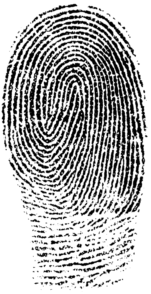
Fingerprinting involves identifying an individual by their unique pattern of arches, loops, and whorls on the ridges of the fingertips. Invisible oils and other biomolecules are laid down on a surface, and crime scene techs visualize those scant traces through a number of methods. Those prints are then compared to a local, national or international database and, hopefully, a match is made and a perpetrator is identified.
But a person’s identity may not be the only thing revealed by his fingerprints, as was recently announced:
- Determining the use of illegal drugs: Researchers from the University of Surrey in England have developed a method to test the residue left in a fingerprint for cocaine using mass spectrometry. More importantly, based on the drug metabolites, it can be determined whether the cocaine was ingested, or whether the suspect simply touched it and traces of the drug remained on his fingertips. New portable mass spectrometers are being developed to make this a technique that can be used in the field at actual crime scenes.
- Fingerprint Molecular Identification (FMI) technology to identify gender, narcotics and nicotine: North Carolina’s ArroGen Group has developed FMI technology, again using mass spectrometry, to identify gender biomarkers, as well as metabolites of nicotine, heroin, methamphetamine, marijuana, temazepam, ecstasy and even some legal medications. This panel of distinctive chemical substances could lead to suspect identification as well as criminal convictions.
- Determining the age of a fingerprint: Researchers at the National Institute of Standards and Technology have developed a method to approximate the age of a fingerprint. This has always been a problem using fingerprints as criminal evidence—a print might prove an individual was in a particular location, or touched a particular object, but was it at the time of a crime, or the week before and therefore possibly insignificant? Scientists have tried to develop a method to date fingerprints based on the breakdown of the biochemical products in the fingerprint, but to no avail. However this method is different and depends on the movement of biomolecules from the ridge to the empty valley sections of the print. Essentially the clearly defined lines in a fresh print will blur and become indistinct with time. How much so will help scientists date an individual print. So far, scientists have been able to distinguish between a day and a week old, a week and a month old, and a month and four months old. This is still a proof-of-concept method, but researchers are working to fine tune the technique, which could be incredibly useful in criminal investigations.
- Determining race of an individual: We’ve previously discussed how to determine race from a victim’s skull, but researchers from North Carolina State University recently announced a technique to determine race from the minutiae of the fingerprint. In a nutshell, there are three levels of examination for a fingerprint. The first level is the one most people are familiar with—those ridge formations called arches, loops, and whorls shown in a standard ink print. The second level is the minutiae—the deviations of those arches, loops, and whorls—where a ridge ends, when it splits in two at a bifurcation, or where the ridge makes a U-turn in a loop or whorl. In comparing those from African-American and European-American backgrounds, researchers found significant differences at the minutiae level, enough to be able to determine from which group the individual came. The study only involved 243 subjects, so these are very preliminary results, but so far the data appears promising.
It is early days so far for many of these techniques, but, with additional study, hopefully they will develop into full-fledged tools for investigators, providing them with more information and hopefully leading to more definitive suspect and victim identifications.
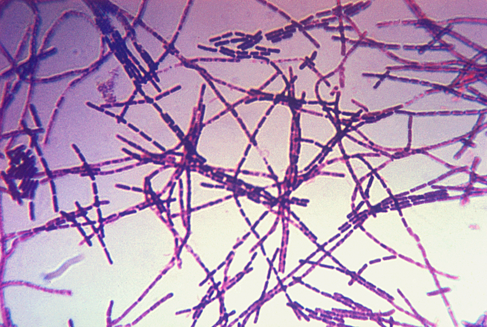
Photo credit: Wikimedia Commons


 17.9%
17.9%