Meet Jess Danna!

We Dannas are an artistic bunch. Long-time readers of the blog may remember the Meet the Dannas! post, that introduced my parents, my two film composer brothers and my languages guru sister. Spoiler alert: that post was written a month before the 2013 Oscar awards where Mychael did indeed win an Oscar for Best Score for Life of Pi.

At the time I mentioned a skilled photographer in the family. Well, that is my oldest daughter Jess, and her time as a professional is just beginning as she is approaching graduation from Sheridan College with a four-year Bachelor of Photography degree. Today is a big day for Jess as she and her graduating class are sharing in their very first gallery showing in downtown Toronto, so I thought it was time to highlight her special skills.


My readers will know her work from the design and photography for the cover for NO ONE SEES ME ‘TIL I FALL (complete with younger daughter Jordan as model, as you will see in an upcoming shot).


She’s also responsible for both my headshot here on Skeleton Keys and my brother Mychael’s headshot over on mychaeldanna.com.

But as a photography student, she’s really been able to stretch her wings from landscapes, to portraits, to advertising mock-ups, to architecture, to compilations. So let’s take a look at some of Jess’s more recent work (the very best way to see them is to click on the first picture for a larger version and to scroll through them; alternatively, you can enlarge individual pictures by clicking on them):

Want to see more? Then check out http://jessdannaphotography.format.com!

It's going to be an interesting few months as Jess graduates and then moves onto bigger and better things, but it will be exciting times for sure. Look out world, here she comes!

Forensics 101: First Archeological Evidence of Buckshot Injuries

Battlefield momument

Battlefield momument

This story is kind of a fun one for me. Not only is it research coming out of my university, but it’s a battlefield site that’s only about 20 minutes from home.

The Battle of Stoney Creek was one of the earlier battles of in the War of 1812 (1812 – 1815). Following the American victory in the Battle of Fort George in Niagara-on-the-Lake, 3,400 American troops camped for the night in Stoney Creek. Even through the British only had 1,600 men, reconnoitering showed them the Americans were badly organized and only thinly sentried with an elongated, broken line of encampment. This was true; in fact, when the battle started, only 1,328 American soldiers out of the total 3,400 were positioned to join the fighting.

Armed with muskets and bayonets, 700 British troops left their camp at 11:30pm, killing the few sentries on duty before moving in to start the battle proper. However, the Americans held the high ground, firing a variation of the traditional ‘buck and ball’ down onto the British, having loaded their muskets with 12 buckshot balls, essentially turning them into shotguns. The Americans held their position and were well on their way to victory when a gap formed in their line, leaving their artillery unprotected and allowing their guns to be taken and their men killed by the British. In fact, the chaos from the lack of light and the uncharacteristic close-quarters fighting led to American officers coming to investigate what they assumed was a commotion produced by their own men. Instead they were taken prisoner by the British. Without direction from their generals, the American soldiers started to wander aimlessly in the dark and many were cut down by their own countrymen. In the confusion, the Americans pulled back to end the battle, unaware they still held both the superior position and number of men. They retreated back to 40 Mile Creek in Grimsby and then finally back across the Niagara River to U.S. soil, never venturing as far into Upper Canada again. The battle only lasted 45 minutes, but by the end, 39 men were dead, 174 were wounded and 152 were captured. Many of the soldiers were quickly buried on site in a mass grave.

In 1899, farmer Allan Smith unearthed human remains and pieces of cloth bearing both the British and American insignias while plowing his land. That area, now called Smith’s Knoll, was finally excavated in 1998 and examined. The excavation revealed 2701 co-mingled skeletal components from 24 individuals. Skeletal remains showed signs of sharp force and projectile trauma, as well as perimortem (at time of death) fractures. In the past, bone injury from musket balls has been well documented, but archeological buckshot injuries had yet to be verified. Whether the dearth of information of this type of injury comes from a lack of evidence (as the British did not use this type of ammunition; it was only used by the Americans) or because there is simply less bone damage and more associated soft tissue damage from buckshot is unknown.

We’ve shown the damage modern bullets can do to bone, but 0.65 caliber musket balls and 0.31 caliber buckshot of the early 19th century were very different: Made of soft lead, projectiles would often become misshapen upon striking the body. Buckshot especially would often become so misshapen, it could penetrate the body, but could not pass through it. As opposed to modern bullets, lead balls and buckshot would only glance off bone, or penetrate enough to become embedded. High-velocity, through-and-through, jacketed ammunition would not exist for another 50 years.

Smith’s Knoll Scapula with buckshot defect.

Smith’s Knoll Scapula with buckshot defect.

Researchers at McMaster University experimented with cloth-encased butchered pork as a substitute for a fleshed human hip in a soldier’s uniform, test firing both the traditional-for-the-time ‘buck and ball’ (a musket ball with 3 smaller buckshot) and buckshot only (with 12 buckshot per cartridge). Their results indicate that some injuries seen in the Smith’s Knoll remains came from buckshot injuries. Instead of the sharply angled, penetrating defects we’re familiar with today, many of the defects were no more than minor depressions, indicative of a low-velocity projectile that has spent most of its energy penetrating cloth, skin and muscle before striking bone. Some bones had multiple defects, clustered close together, indicating buckshot fire from close range, not allowing the buckshot to separate as it left the musket and flew through the air. Due to the known history of the battle, it is impossible to tell if the skeletal remains are those of British soldiers cut down by American militia, or militiamen felled by friendly fire.

The War of 1812 is a curious thing. It went on for nearly three years, and is considered to this day by Britain to be a minor part of the Napoleonic Wars. The British torched the White House in 1814 and kept the Americans at bay during a number of decisive battles in Southern Ontario (does anyone but a Canadian know the name ‘Laura Secord’?) avoiding being annexed to the United States, but didn’t fare well in fighting in New Orleans or Baltimore. By 1815, when the Treaty of Ghent was signed to bring an end to hostilities, nearly 20,000 men were dead, a military stalemate was called, and the borders remained exactly where they were. However, due to the lack of clear winners or losers, no bad feelings persisted and friendly trade immediately resumed.

Photo credit: Wikimedia commons by Nhl4hamilton and L. Lockau et al

Disease Resistance: Could You Be A Mutant?

It’s a story right out of one of the X-Men movies—people who are born with genetic differences that give them an evolutionary advantage over their fellows. In this case, that advantage is over catastrophic disease.

We’re all familiar with how most disease research is accomplished: scientists study those who are afflicted with a particular disease to see how it affects them overall and how it affects their individual biological systems. But Dr. Stephen Friend of Sage Bionetworks in Seattle led a team of scientists from New York’s Icahn School of Medicine at Mount Sinai on a different tack—to study individuals who should suffer from or should have died from disease, but never became sick in the first place. To do this, they examined the genetic profiles of nearly 600,000 subjects from 12 previous genetic studies and looked closely at 584 Mendelian diseases (diseases caused by single gene mutations that are inherited according to Gregor Mendel’s Laws).

Mendelian genetics is the type of genetics most high school students studied in biology class. Remember Punnett squares? That’s Mendelian genetics—gene traits that are dictated by the alleles of a single gene. Let’s review and look at a very simplified version of eye colour. If a parent has brown eyes, they are expressing the dominant allele of the eye colour gene, which is brown (B). But everyone has two copies of that gene, one donated from their mother and one from their father. Because of the dominant brown allele (B), the second allele could be a second dominant brown allele (B) or a recessive blue allele (b) masked by the dominant brown. In high school biology terms, this parent could be BB, or Bb; both allele pairs would result in brown eyes. Only a parent with blue eyes is guaranteed to be bb, because two recessive genes will allow the recessive colour to show instead of being masked by the dominant colour. But if you pair two brown eyed parents, both of whom come from a brown eyed and blue eyed parent themselves, you will have two parents who are Bb and the below Punnett square shows what their offspring statistically should look like.

This is my husband and I exactly. Both ours mother have brown eyes, both fathers have or had blue eyes, but we both have brown eyes, therefore, we must both be Bb. We have two daughters, one with green eyes (genetically blue in this simple example; the green colour comes from additional masking gene alleles) and one with brown eyes.

Mendelian diseases are similarly governed by a single mutation on a single gene. Some of the diseases they looked at were Cystic Fibrosis (a disorder of the exocrine glands that affects the lungs, pancreas, intestine, liver, and kidneys), Smith-Lemli-Opitz syndrome (a mutation in the cholesterol pathway which leads to significant developmental delays or even death), epidermolysis bullosa (a devastating blistering skin condition), familial dysautonomia (a disorder of the nervous system), and Pfeiffer syndrome (which causes the bones in the skull to fuse early, leading to a misshapen head, bulging eyes and abnormal brain development). And what researchers found both surprised and delighted them. Of the 589,306 subjects tested, looking at 874 genes that guarantee the development of disease, they found 13 individuals who genetically had one of the mutations and yet had no indications of disease. For all intents and purposes, they are resistant.

Yes, I hear you cry: 13 people out of 589,306? That’s only 0.0022%, so what’s the big deal? The big deal is the answers these people may hold. Why aren’t they sick? What is it about their genetic makeup that counteracts a devastating mutation that might have already killed them otherwise? Discovering those secrets could potentially help the 70,000 people worldwide living with Cystic Fibrosis, desperately waiting for lung transplants while fighting trying to take their next breath. It could help the more than 500,000 people worldwide who suffer from epidermolysis bullosa, whose skin blisters and peels off at the lightest touch, and who can die from cancer or from infection of the exposed derma.

It's clear that 13 subjects are not enough to power any kind of real scientific study, but they are a promising start. Scientists now hope to recruit healthy volunteers that are willing to share their genetic information. Interested? Then the Reliance Project may be for you. Stop by the site, and take a look and join me in signing up. As they say at the Reliance Project: ‘Join the search. Be a hero.’ You might just save a life along the way.

Photo credit – Wikimedia Commons

A Second Viking Settlement Discovered in North America

Newfoundland coastline

Newfoundland coastline

Last week, we talked about the science of space archeology, and how, with the help of high resolution satellite scans taken 400 miles above the earth, a well-trained and intuitive eye can discern archeological sites hidden on the earth’s surface. This week, we’re going to talk about Dr. Sarah Parcak’s latest amazing discovery based on this technology.

The Vikings were a group of Scandinavians, first known for their international trading, but later known universally for their violent raiding of other lands and cultures. They were groundbreaking nautical engineers for the time and their carefully crafted vessels allowed them to travel from present day Norway, Sweden, and Denmark to Britain and Scotland, Russia, Turkey, Iran, and even all the way to North America.

The Vikings used water to move between locations, be it rivers to move through Europe proper or the Atlantic Ocean and Mediterranean Sea to go further. Starting in the 8th century, they set out to trade with peoples in other lands, but quickly learned that it was more successful and lucrative to simply raid those lands instead. They spread across the north Atlantic, moving from Scotland to Iceland, and then to Greenland.

But how did they so successfully set off into the unknown, and not be lost forever at sea? The Vikings were not only expert naval engineers, but were also expert navigators. They learned how to be able to detect land up to 50 or 60 miles away, simply by watching distant clouds, identifying sea birds out for a day of fishing, or by the smell of grass and other plants, carried by the strong sea breezes.

One of the great Viking explorers was Erik Thorvaldsson, better known as Erik the Red. Erik was part of an Icelandic settlement before he was exiled for three years in 982 A.D. after he murdered several members of the settlement. He and his men set sail, only to discover Greenland. It was Erik’s son, Leif Erikson, who is credited with the discovery of North America. He and his men sailed from Greenland, but were caught in a storm and driven due west, where they discovered a previously unknown coastline. Scholars studied the Viking texts of the time and believe Erikson found Baffin Island in Canada. From there he sailed south, down to Labrador, and then to Newfoundland.

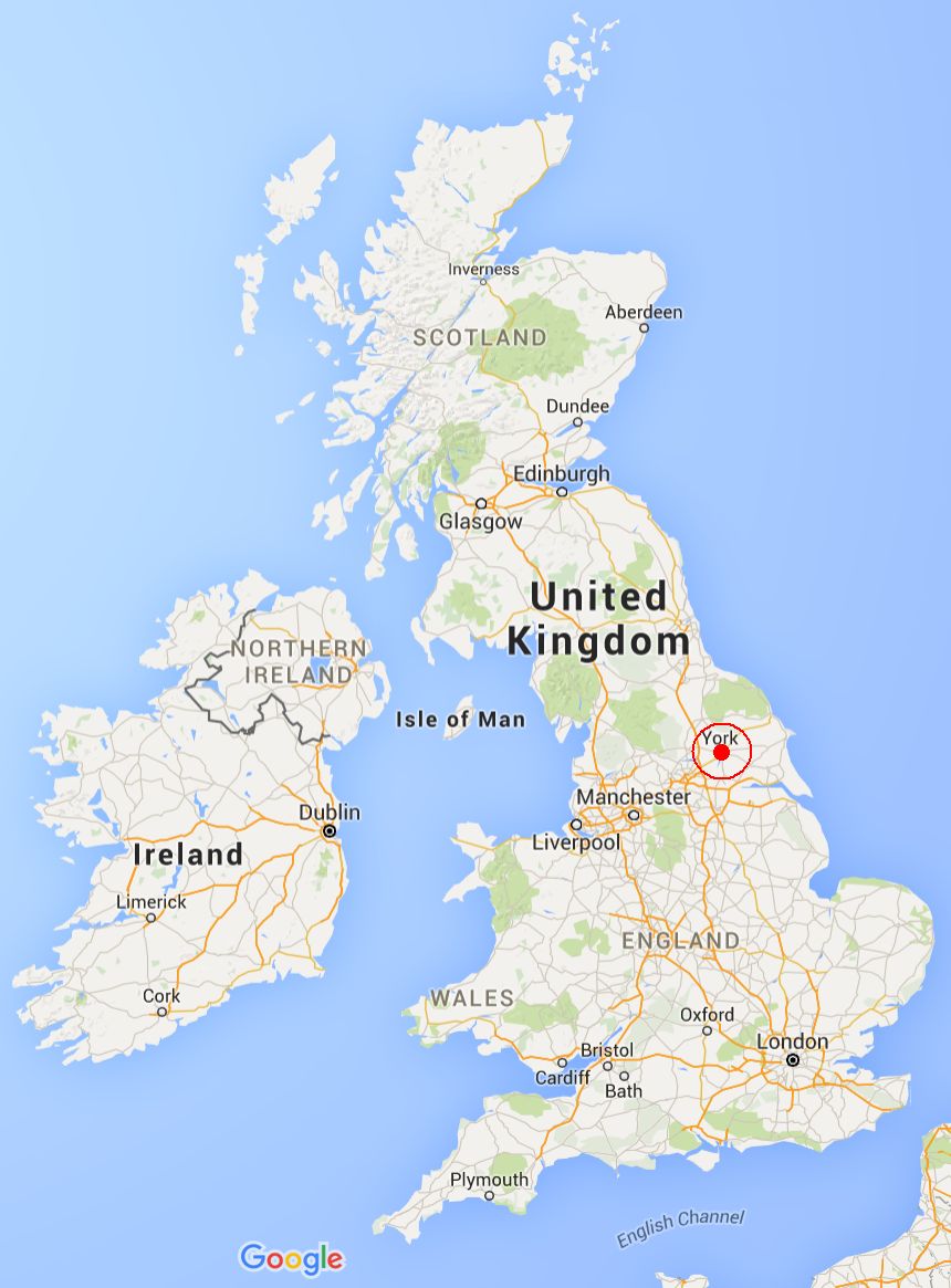
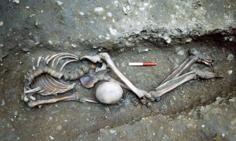
In 1960, the only officially designated North American Viking settlement was discovered in northern Newfoundland. Archeologists called it L’Anse Aux Meadows, and found there the foundations of traditional Viking longhouses for approximately 90 people, and traces of metal works, a technology native Canadians did not have at the time. But archeologists also found something they could not explain—seeds from a plant that only grew hundreds of kilometers south of that site, leading them to believe that there must have been at least one other settlement south of L’Anse Aux Meadows.

Dr. Sarah Parcak, an archeologist at University of Alabama at Birmingham and the director of the Laboratory for Global Observation, took on the challenge of trying to find this new settlement. Using the Worldview satellite that resolves images down to 10”, she examined the Newfoundland coastline. To do this, she studied near infrared scans after processing them for false colour to pick up differences in the vegetation—indications of decreased health of plants visible in the near infrared frequency could indicate the presence of foundations or other man-made objects below the surface.

Dr. Parcak found a potential site at Point Rosee, Newfoundland. Located on the west-facing coast on the southern edge of Newfoundland, it lies 370 miles southwest from L’Anse Aux Meadows. Among other possible structures, she identified a rectilinear shape with the same dimensions as a longhouse found at L’Anse Aux Meadows. This was a very strong indication that Point Rosee could be a related archeological site.

The first step for Dr. Parcak’s team was to do a non-invasive survey of the area, which they accomplished using a magnetomer to detect subtle differences in magnetism of the scanned soil. The magnetomer will not only  indicate the presence of metals but it will also provide evidence of burning or soil disturbances. A number of ‘hot spots’ were discovered and compared to the satellite  scans. It was this successful comparison that earned the team a two week test excavation to see if the they could find any physical evidence of a Viking settlement.

After surveying and gridding off the area to match the terrain exactly to the satellite images, the team dug several test trenches. It was backbreaking, muddy work, but they discovered several items of interest: some seeds, possible fragments of metal work, some clumps of what the team thought might be slag (a byproduct of metal work and a significant indicator of Viking activity), and darkly striped soil seen before in other Viking sites from their use of slabs of turf to insulate buildings.

In the end, the seed was determined to be from the 17th century and archeologists had to admit it could have filtered down into the extremely moist soil hundreds of years later. But it was the suspected slag that ended up as the crucial evidence. While it wasn’t actually slag—it was fire roasted bog ore—it represents a step in preparing ore for metalwork and is indicative of the presence of a group of people with skills different from the resident aboriginals of the time.

It is this single piece of evidence that will now set the course for future excavations. It is a very strong possibility that a Viking settlement occupied Point Rosee over a millennia ago, a full 500 years before Columbus sailed west to ‘discover’ a land found and temporarily settled by other Europeans centuries before. Dr. Parcak and her team hope to shine a light on a time period that is still mostly a mystery, expanding on the history of a new land, as well as the saga of a lost people.

Photo credit: Kenny Louie

Forensics 101: Space Archeology?

A new term came to my attention last week, one that on first glance seems a bit of a misnomer. It came tied to a really neat story, so we’re going to look at the scientific field this week—what these scientists do and what their work tells them—and then we’ll explore their groundbreaking discovery next week.

The term is ‘space archeology’ or, alternatively, ‘remote sensing techniques in archaeology’ (though I think we’d all agree ‘space archeology’ is WAY cooler). At first glance, one would think this is simply archeology in space, except we’re nowhere near having the skills or technology to do that. So what is space archeology? In the end, the answer is quite clever: it’s using satellite scans of the earth taken from space to identify previously undiscovered archeological sites.

The way space archeologists do their work is quite ingenious. Visible light scans of the planet’s surface may show absolutely nothing. But when infrared scans are used after being processed using false colour, chemical changes to the landscape caused by building materials and the activities of ancient civilizations are revealed. NASA’s high resolution scans are used as the raw data for the analysis, allowing scientists to discern subtle variations in the earth’s topography. The key to this analysis is grounded in vegetation—plants that live on top of stone are simply less healthy and will have reduced levels of chlorophyll. Find the unhealthy plants, and you may be well on your way to finding the site of an ancient civilization.

Enter Dr. Sarah Parcak, an archeologist at University of Alabama at Birmingham. She is responsible for the discovery of several amazing archeological sites, many of them lost for centuries or even millennia. For instance, the picture above shows an infrared scan of what looks to the naked eye like a patch of desert. But move out of the frequency of visible light and into infrared, and suddenly a network of city streets and buildings are revealed. It’s an amazing look at what hides below the earth’s surface. Which city is this? Tanis, the historical city used in Raiders of the Lost Ark, lost for over three millennia to the sands of time. So far only a small trial area has been excavated, but mud-brick structures were discovered a foot below the modern surface.

Next week we’ll be back to talk about an amazing discovery that could rewrite the early history of North America. See you then…

Photo credit: University of Alabama at Birmingham

Forensic Case Files: The Bard’s Missing Skull

There has been controversy for years about who Shakespeare really was. History tells us he was the son of a glove maker, born in Stratford-upon-Avon in England in 1564, who grew up to be an actor, poet and playwright. But doubts were raised that someone born in a small village and living so far outside royal life would be able to write about it so eloquently, and some have proposed that Sir Francis Bacon or Christopher Marlowe were actually ‘The Bard’. But the man most recognize as the ‘greatest writer in the English language’ is known to have died four hundred years ago on April 23, 1616. He was laid to rest two days later in the chancel of Holy Trinity Church in his beloved Stratford-upon-Avon. Later, his wife, daughter and son-in-law were buried beside him.

Two stories of a strange grave robbing surfaced roughly 250 years later, in 1879 and 1884. They describe a doctor digging up Shakespeare’s head in 1769, possibly to sell to an art dealer. There is a theory from the time that a person’s genius could be discerned from their skull alone, so Shakespeare’s skull would have had significant worth.

Recently, that tale was put to the test as researchers from Staffordshire University were allowed to come into Holy Trinity Church with ground penetrating radar equipment to scan the grave under an etched stone slab. And what they found supports those stories—the results show a disturbance at the head end of the grave showing where dirt was removed and replaced, and the skull does not appear to be present. The scans also show that Shakespeare and his family were not buried in coffins, but simply wrapped in cloth shrouds and entombed in shallow graves, which would have certainly made grave robbing an easier task.

Researchers realize their results ask more questions than they answer, but they are determined to go back to the records of the time to try and solve the mystery of Shakespeare’s missing skull. Was it truly stolen, or could it reside in another church or in a family member’s tomb instead?

An interesting side note to the theft is the epitaph chiseled on Shakespeare’s tomb, one the robbers most certainly ignored:

Good friend for Jesus sake forbeare,

To dig the dust enclosed here.

Blessed be the man that spares these stones,

And cursed be he that moves my bones.


Photo credit:  Steve

Queen Nefertiti’s Tomb Discovered?

We love interesting burials here at Skeleton Keys. Add in some fascinating history and we’re in heaven. We spent lots of time examining the process of finding and identifying Richard III. We covered the potential discovery of the remains of Joan of Arc and King Alfred. We looked at the plague burials of London, the Roman burials of York, and even those entombed at Pearl Harbor. But we’ve never tackled ancient Egypt.

This past week, there was a press conference in Cairo. The Egyptian minister of antiquities announced that new research being conducted in King Tut’s tomb revealed the possible presence of two hidden chambers off the room that held the royal sarcophagus. Seeing as almost everyone thought the tomb had been fully excavated by Howard Carter between 1922 and 1932, the announcement came as quite a surprise.

The one man who thought there was more to discover was Dr. Nicholas Reeves, an archeologist from the University of Arizona. Known as a scientist who often makes discoveries by fully analyzing the research of others, he published a paper in 2015 based on radar scans of the tomb done by a team of conservators from Factum Arte. Factum Arte’s scans were commissioned in order to produce a replica of the tomb for an Egyptian project to preserve tombs in the Valley of the Kings. After the scans were complete, they were uploaded to the Internet for all to see. When Dr. Reeves examined the highly detailed scans, he saw what everyone else had missed—the outline of two doorways in clear, straight lines beneath layers of plaster and paint. His analysis of the data prompted further scans to determine if there was any truth behind his theory.

Dr.  Hirokatsu Watanabe, a well-known Japanese radar expert, was brought in to run fresh scans of the north and west walls of the tomb. Immediately following the scans, Watanabe was “90% positive” of the presence of a hidden room behind the north wall. Six months later, the full analysis is complete and Watanabe reported finding empty space on the other side of both walls, with metal and organic material behind the north wall, and organic material behind the west wall. Organic material in this case could be anything from wooden items to human remains.

The quality of the plaster in those two areas is also different from the tomb walls themselves. It is composed of a softer, grittier material than the tomb walls proper. More specifically, this type of gritty plaster exactly matches the material sealing another door opened by Howard Carter in his initial excavation. Carter kept some of the material so Reeves was able to do a direct comparison.

Reeves has a theory about the reported organic materials. It is his opinion that the remains of Queen Nefertiti, stepmother to King Tut, are buried in the chamber behind the north wall. If so, KV62, the most important archeological find of the 20th century, may prove to also be the most important archeological find of the 21st century.

Howard Carter was an amazingly thorough archeologist and painstakingly took a decade to document and excavate King Tut’s tomb, a dedication that was considered unusual for the times. But imagine such an excavation with modern archeological and scientific tools at the researchers’ disposal. They would be able to do field testing radically unlike anything Carter could achieve and in-lab testing well beyond that. Not to mention that current conservation skills far outstrip any of a century ago, ensuring the safety of any objects inside the tomb, sealed away for the past 3,339 years.

So what are the next steps? National Geographic has been invited to send in a team of specialists to confirm the previous radar data as well as to determine the thickness of the walls. Once that is determined, Dr. Reeves would like to use a tiny fiber optic camera to breach the wall with as little damage as possible to visualize any open space beyond without defacing the beautiful painted murals that cover both the north and west walls. He would also like to talk to Japanese conservators with experience in removing wall paintings intact in case they have to fully breach the wall to excavate behind it.

It’s a very exciting prospect, but one that will take time and care to achieve. So patience will be the watchword as the Egyptian government carefully oversees the entire process. But they will not be the only ones to pay attention. To say the whole world will be watching is surely not an overstatement.

Photo credit: By Philip Pikart - Own work, CC BY-SA 3.0

The Bugs Are Going To Win… Or not?

First of all, our apologies for being AWOL for the last several weeks. We were on deadline on the developmental edit of LONE WOLF and barely had time to sleep, let alone come up with a well-researched article for the blog. But the manuscript is done now, and handed in, and we’re back in business!


Normal
0


false
false
false

EN-US
X-NONE
X-NONE


 <w:LatentStyles DefLockedState="false" DefUnhideWhenUsed="true"
DefSemiHidden="true" DefQFormat="false" DefPriority="99"
LatentStyleCount="267…

A T4 bacteriophage poised to infect a bacterium.

I have a saying that I seem to repeat often: “The bugs are going to win.” This refers to the oncoming crisis humankind is likely to have against microbes (or ‘bugs’ as we generically refer to bacteria in the lab). In a day and age when factory-farmed animals are given antibiotics prophylactically and too many medical professionals still consider knee-jerk prescription writing for antibiotics an acceptable act, news stories repeatedly warn of the number of strains of bacteria resistant to all antibiotics. Another continual theme are reports of now-resistant bacteria that used to be susceptible to a shrinking number of heavy hitting antibiotics used in only the most severe hospital cases.

In many ways, the thought of these tiny organisms taking out human kind is hard to wrap your brain around. We’re the top of the food chain; we have technology and science on our side. How can they top that? Part of the answer is very simple—replication rate. Your average bacteria divides into two identical daughter cells approximately every 20 minutes. It takes humans approximately 20 years for that same replication. This rate of bacterial replication leads to a very large number of progeny—in one day, a single bacterium can produce 4.7 x 10^21 bacteria. Even if the mutation rate is only 1 in 10^10 DNA base pairs, depending on the genome size of the bacteria, hundreds of mutations can occur in a span of less than 10 hours. Some of these mutations could lead to an inactive bacterium, but one of them might lead to a new gene conferring resistance to one of our last ditch antibiotics.

What happens when we can no longer control bacteria with a pill or injection and we enter the post-antibiotic age? It will be like going back to the early 20th century, before the advent of penicillin. Pneumonia would be a fatal infection. Bacterial epidemics similar to the Black plague that killed 30 million people—a full third of Europe at the time—could once again be a scenario faced by the world. Those who are very young and very old will be at risk due to compromised or immature immune systems. It’s a very scary concept.

So how can we beat the bugs?

  • Develop new chemical therapies or multi-therapies: Scientists at Merck Research Laboratories in New Jersey recently announced two separate compounds, tarocin A and tarocin B, that makes Methicillin-resistant Staphylococcus aureaus (MRSA; a bacteria well known for its resistance) susceptible to antibiotics again. Tarocins A and B target the bacterial cell wall, and, when used with standard antibiotics, kill the bacteria effectively in mice. Human trials will be the next test.
  • Find new natural anti-bacterial compounds: Dr. Gerry Wright at McMaster University has been sending students around North America to sample soil in different areas. Compounds from those soils are then tested against bacteria. If the bacteria are killed, the samples are then further tested to determine the successful compound. So far the team has made two notable discoveries:
    •  AMA, a molecule produced by a fungus in Nova Scotia soil that is able to knock out one of the strongest and most worrisome antibiotic resistance genes, NDM-1. NDM-1 is such a concern that the World Health Organization identifies it as a global health threat. When AMA is used, regular antibiotics once again become effective.
    • Teixobactin is an antimicrobial molecule produced by one bacteria to kill others and was discovered in the soil from a field in Maine. The compound has been shown to be effective in killing multiple antibiotic resistant strains of bacteria.
  • Revisit OLD remedies: A one thousand year-old Anglo Saxon recipe to treat eye infections using onion, garlic, wine and cow bile has been found by modern scientists to kill MRSA. Rather than being any individual ingredient that was effective, it was the combined recipe that killed 90% of the MRSA in the sample.
  • Use their own natural enemies against them: We’re all familiar with the concept of viruses. Bacteriophage are viruses that attack bacteria (see the above photo), acting as all viruses do—they inject their contents into the bacterium and highjack it’s replication machinery to produce hundreds or thousands of copies of itself, eventually bursting the bacterium, killing it. Scientists in Russia and parts of Europe have used bacteriophage for nearly a century as antibacterials. The Western world is now looking at phage therapy as a potential replacement for antibiotics.

In the end, the question remains can we beat the bugs, and that remains to be seen. In the meantime, be conscientious citizens of the world. Do not demand antibiotics for your kids because they have ear infections, of which a full 70% can be caused by viruses; wait and let the infection run its course and it will likely clear up just as fast without antibiotic usage. Don’t run to the doctor every time you have a cold until you hit the 7 to 10 day range and a secondary infection may have set in. We should only use antibiotics when they are truly necessary. And maybe, just maybe, the bugs won’t win after all.

Photo credit: Xvivo Scientific Animation

The Women of Stonehenge

Stonehenge—the ancient Neolithic monument in Wiltshire, England—was built approximately 4,000 – 5,000 years ago. The modern configuration of the henge was built over a span of nearly 2,000 years—from the initial shaping of the land, to construction of a timber structure, the transition to the first stone structure, and finally to the transport and configuration of the massive twenty-five ton sandstone blocks and their associated blue stones that remain on-site today.

Stonehenge was built and used during a time before history was documented in written records. While word of mouth passed down stories for generations, this many millennia after its creation, some of the purposes of Stonehenge have been lost to the ages. However, modern research proposes a number of uses for the site, including that it may be one of the oldest recorded organized burial grounds of both human and animal remains. It also served as an astronomical calendar, arranged to align with sunset at the winter solstice and sunrise at the summer solstice with a precision that is almost unbelievable considering the knowledge and tools of the time. It was used for religious ceremonies, and it has also been suggested Stonehenge was used as a place of healing based on the condition and associated traumas of the remains discovered on site.

Computer rendering of the completed Stonehenge

Computer rendering of the completed Stonehenge

Stone Age human remains were recovered from Stonehenge in 2008 and have been studied by researchers at University College London. The original find revealed over two hundred cremated individuals in a chalk pit. From this, fourteen females and nine males were definitively identified using CT scans and radiocarbon dating to determine not only the sex but the age and date of burial of the remains.

It was the proportion of females to males that most impacted researchers. In today’s society where North American women still experience the resistance of the glass ceiling and many women internationally are simply fighting for autonomy and the right to vote, researchers were impressed by the clear acceptance of women in positions of power. Only the most influential members of society would be buried in such an important spiritual place, so this is a clear indication of a gender-equal society a full five thousand years ago.

In many modern depictions of Stonehenge’s history, ritual and rite are only conducted for men and by men, but science shows that Stone Age society was perhaps more advanced than we previously believed. Sometimes it’s good to look to the past to inform our modern lives, and this might be a good example of when lessons can be learned from those who have gone before us.

Photo credit: Peter Trimming and Wikimedia Commons

It’s Cover Reveal Time!!

Having a new book release is fun, but it’s fun in stages: The satisfaction of typing ‘The End’ on the first draft. The joy of signing a contract (after all the uncertainly of drafting and convincing yourself that the whole story is garbage, it’s like that moment when Sally Field won the Oscar for Places in the Heart.)

Holding an advance copy in your hands for the first time, and likewise the final copy. Seeing your book on bookstore shelves. All that hard work, finally realized.

Part of this process is getting the cover. It lands in your inbox and you have a brief moment of ‘what if I don’t love it?’. You open the image peeking through your fingers. And then you realize that people more talented than you, people who really know what they are doing, are on your side and they’ve made something fantastic for you.

That’s how we felt we received the cover for LONE WOLF, the first book in the new FBI K-9 thriller series we're writing as Sara Driscoll. And we’re thrilled that we can finally show it off. We can’t wait to see this on shelves in December!

When a madman goes on a bombing spree, an FBI K-9 team of one woman and her dog is the key to stopping him before more innocents die and panic sweeps the Eastern seaboard.

Meg Jennings and her Labrador, Hawk, are one of the FBI’s top K-9 teams certified for tracking and search and rescue. When a bomb rips apart a government building on the National Mall in Washington D.C., it will take all the team’s skill to locate and save the workers and children buried beneath the rubble.

More victims die and fear rises as the unseen bomber continues his reign of terror, striking additional targets, ruthlessly bent on pursuing a personal agenda of retribution. Meg and Hawk join the task force dedicated to following the trail of death and destruction to stop the killer. But when the attacks spiral wide and no single location seems safe any longer, it will come down to a battle of wits and survival skills between Meg, Hawk, and the bomber they’re tracking. Can they stop him before he brings the nation to the brink of chaos?


Isn’t that eye-catching and atmospheric? We love that Hawk is right out front because we have no story without him.

Hit the comments and let us know what you think!

Zika: Emergence of a New Epidemic

Electron micrograph of Zika virus particles.

Electron micrograph of Zika virus particles.

You’d have to be living under a rock for the last few weeks to have missed the fact that a new virus—called Zika—has been making headlines. In reality, Zika is not a new virus at all, however its effects in the last six months in Brazil and other south American countries has been utterly alarming. Worst, one of its most terrible of its outcomes strikes the most fragile of us—unborn infants.

The Zika virus was discovered in 1947 in the Zika Forest in Uganda. It is one of a number of viruses in the Flaviviridae family which includes dengue virus and West Nile virus. By far, the main method of transmission is via the mosquito Aedes aegypti, therefore the areas of mosquito-borne Zika infection follow the natural distribution of Aedes aegypti in an equatorial band around the planet. Historically, disease caused by Zika has been considered mild and mostly trivial with four out of five infected people never knowing they were ever infected. Those who develop symptoms usually present with a low grade fever, joint and muscle pain, and headaches lasting from approximately two to seven days. Since there is no effective medicine or vaccine, symptoms are treated directly—rest, plenty of fluids, and acetominophen to ease the aches and pains. For most patients that is sufficient, and severe disease and death were extremely rare.

However, a disturbing picture emerged from Brazil last November when the news broke that 4,000 babies had been born in 2015 with microcephaly—a birth defect in newborns with abnormally small heads and incomplete brain development. Compared to the normal average of 150 babies born with microcephaly in a single year, this is a staggering increase. While we’re still waiting for definitive evidence that this increase is due to the Zika virus, Zika was first identified in the country in May of 2015, and some pregnant women at the time showed evidence of the virus’ RNA genome, leading public health officials to infer that Zika was responsible. Late last week, Columbia reported three people who died of Guillain-Barré syndrome—a disorder where the body’s immune system attacks the peripheral nerves causing muscle weakness and paralysis—following Zika infection.

Recently, the World Health Organization took the significant step of defining Zika as a ‘Public Health Emergency of International Concern’. However, it is difficult to make recommendations about Zika since the virus is not well studied. Less than 200 scientific papers mention Zika, only a handful of which are notable for significant information. And the fact that so much is still unknown—all methods of transmission, life cycle of the virus, where it replicates inside the body, and what body fluids it can be found in and for how long—ties the hands of public health officials who are trying their best to prepare people on how best to protect themselves. The virus has been found in saliva, blood, semen, and urine, leading to warnings about sexual transmission, pregnant woman kissing anyone other than their partner, and to blood banks setting restrictions on blood donations in a window following travel to Zika-ravaged countries. Some countries, like El Salvador, have even suggested to women that they refrain from getting pregnant for the next two years, a difficult issue in a Catholic country where most methods of birth control are forbidden. At this point, the main preventative strategy is to avoid travel in the twenty-six countries with documented Zika infections. If not, avoidance of mosquito bites is the next best thing, by using bug spray, wearing long sleeves and pants to cover the skin, and sleeping in rooms with window screens or air-conditioning.

This story has struck a personal note in my day job as a manager of an infectious diseases lab. Flaviviruses are one of our main areas of study, with past projects including major NIH studies of both West Nile and dengue fever. In fact, dengue virus is carried by the same mosquito as Zika, opening up the possibility of co-infection or super infection (infection of a second virus following infection of the first). While the West Nile study was of North American cases, the more recent dengue fever study involved cases and controls from Central and South America and southeast Asia. Currently, we’re looking at the tens of thousands of samples in our freezers from that study and wondering how many of them might also contain Zika. Discussions are already well under way and we hope to join the fight against Zika very soon. We’re certainly better situated to hit the ground running than most labs considering our existing sample bank, and we have high hopes that we can make a meaningful contribution.

So where do we go from here? First and foremost, remember all the public health agencies are doing their best to keep people safe. It’s a damned if you do and damned if you don’t situation—people will be upset if recommended precautions seem too severe, or if they turn out to be too lax as additional information is discovered.  They’re doing their very best based on extremely limited information. And while for the majority of people, Zika is not likely to be a major health concern, for the sake of those that are more significantly affected, it behooves us all to be diligent to protect the vulnerable.

Photo credit: Wikimedia Commons

Debunking JFK’s Single Shooter Theory?

On November 22, 1963, President John F. Kennedy was fatally shot in Dealey Plaza in Dallas, Texas, as he rode in a motorcade on the way to a luncheon at the Dallas Trade Mart where he was to meet with the city’s top leaders and businessmen. In 1963, the science of forensics was in its infancy. There was incredible outside pressure upon law enforcement to identify and apprehend the guilty party, giving a grieving country closure and hopefully allowing it to move on with Lyndon B. Johnson at the helm. Lee Harvey Oswald seemed to fit the bill as the perpetrator of the horrific crime, only his shooting by Jack Ruby closed the case with even more speed than investigators ever anticipated.

There have been multiple investigations of the JFK assassination. The first was the Warren Commission, meeting from December 1963 to September 1964, which concluded that Oswald had acted alone. From this investigation, the ‘Single Bullet’ theory (named the ‘Magic Bullet’ theory by critics) was proposed: a single bullet, fired from Oswald’s rifle from the sixth floor of the Texas Book Depository, struck President Kennedy in the back before passing through his neck to then strike Texas Governor Connally in the back, shattering his fifth rib before passing through his right wrist to finally lodge in his left thigh. Early critical analysis rejected this theory, stating that the bullet would have to change direction in mid-air several times to fit the documented injuries. However, current forensics and materials knowledge has confirmed that with a slight change in position of the two victims, a single bullet could realistically explain the pattern of injury. However, Kennedy would have survived that shot. But a second bullet struck the back of Kennedy’s head, shattering the skull and fragmenting inside the brain, leaving him immediately brain dead and clinically dead within thirty minutes.

Despite the Warren Commission decision, theories of a second gunman on the grassy knoll persisted. In 1979, the United States House Select Committee on Assassinations ruled that JFK and Governor Connally’s injuries were caused only by Oswald’s two shots, however audio analysis indicated there could have been a second shooter, fanning the flames of conspiracy theorists who insisted that Oswald hadn’t acted alone.

Five bullet fragments were recovered during the original investigation and were analysed to determine if they might have come from more than the two bullets that struck the President. In 1978, radiochemist Dr. Vincent P. Guinn was allowed to test the fragments using Neutron Activation Analysis—a procedure to test trace elements in a material without destroying the sample. From the analysis of the trace elements antimony, arsenic, copper, and silver, Guinn determined that all the fragments came from the same two bullets with an identical composition.

Dr. Clifford Spiegelman of Texas A&M University used modern statistical techniques to re-examine those same five bullet fragments. And his findings directly contradicted those of Dr. Guinn. As Dr. Spiegelman explains, there were several significant problems with Guinn’s analysis. The first was the belief that a single chemical test, as opposed to a multi-disciplinary analysis, would be sufficient to yield reliable results. The second problem was the assumption that trace elements during single manufacturing run of the material would be consistent throughout the production line. 

Idealized element composition during manufacturing.

Idealized element composition during manufacturing.

In terms of ammunition, Dr. Guinn and the FBI assumed that bullets with the same composition would have come from the same box. Instead, Dr. Spiegelman found the actual production of the sample was anything but consistent:

Actual antimony sampling during multiple bullet production runs.

Actual antimony sampling during multiple bullet production runs.

This means that the original hypothesis behind the chemical analysis of the JFK assassination bullet was fundamentally flawed. To demonstrate this, the team from A&M analyzed thirty bullets to determine their trace element profile. Of the thirty, one of the modern bullets actually matched the profile of the JFK bullet fragments, further proving how a simple chemical analysis is insufficient to prove a chemical profile so unique to confirm it could be the only bullet involved in an assassination.

Dr. Spiegelman is adamant that this does not mean there was a second shooter in Dealey Plaza. But he does insist Dr. Guinn’s original assessment that there were only two bullets and they came from the same box of bullets and the same rifle has to now be considered totally inconclusive.

Photo credit: Wikimedia Commons and Dr. Clifford Spiegelman, Texas A&M University

Ancient York Cemetery Sheds Light on the Roman Empire

Located approximately 300km north of London, the city of York was a major Roman outpost close to the northern border of their British domain. The area had been populated since approximately 7000 B.C., but the city itself was founded as Eboracum in 71 A.D. when it became a Roman fortress and settlement. For hundreds of years, the city was presided over by a series of emperors. However, after the fall of Rome, the city was overrun by the Angles in the 5th Century.

An ancient Roman cemetery was discovered in 2004 in the gardens of Driffield Terrace as they prepared to develop the property. The York Archeological Trust excavated the site and was more than a little surprised to find 80 sets of remains from the Roman period dating from the early second century to the late fourth century A.D. The cemetery is located on what would have been the outskirts of Eboracum, across the river from the Roman fort.

The remains were determined to be those of Roman gladiators based on several details. The bone structure of the individuals indicated that they were all men of less than forty-five years old and of large stature with heavy muscle attachment points, indicating a muscular physique. Remodeled bone told the tale of significant battle trauma and one set of remains even showed signs of a large animal bite, likely a lion or bear from the gladiatorial ring.

In 2010 testing was done on some of the bones based on strontium isotope analysis and it was determined that the individuals studied likely came from diverse areas and were not all of British origin. Of the eighteen individuals tested, only five came from York. The remaining thirteen came from outlying areas of Britain, mainland Europe and the parts of the Mediterranean, and the Middle East. Traces of carbon and nitrogen in the bones also led the team to the conclusion the gladiators ate a very different diet than the majority of the population of York, confirming the theory that they came from different geographical origins. A number of the skeletons had been decapitated and the skulls buried with them on their chest, between their legs, or at their feet.

However, at the time, scientific techniques to identify the exact origins of the men didn’t exist. But current day cutting-edge genome wide analysis now allows for the level of precision and analysis required to identify genealogical locations. Researchers from Trinity College Dublin selected seven skeletons for testing. Of these seven, six were found to be of British origin and related to the modern Welsh people, suggesting a migration from the area with the arrival of the Angles in the fifth century. The remaining skeleton however was radically different, and researchers matched his DNA sequences to the Middle East, specifically to Palestine, Jordan or Syria.

This is the first definitive evidence of the scope of the Roman Empire and the movement of troops within it. In a time where mobility of troops was an enormous proposition, it is clear that some of the centurions were very well travelled. The study also confirms the multi-ethnic composition of the Roman Empire.

Photo credit: York Archeological Trust

How an Astonishing Evolutionary Trick Could Lead to a New Cancer Treatment

This post might be a little inside baseball and angled toward the science crowd, but I ran across a very interesting story this past week that I wanted to share. It had to do with how a surprisingly small change in DNA sequences in the earliest single-celled organisms not only lead to multicellular organisms, but to the diversity brought on by sexual reproduction, and could possibly even lead to a new treatment to one of mankind’s worst diseases, cancer.

We all know that life evolved on this planet, first from individual molecules, to organized clusters, to single-celled organisms, and finally to multi-celled organisms. For anyone who sat through science class, this is simply an accepted truth. But the enormity of that truth got a little wake-up call in a paper, recently released in the on-line journal eLife. Given the fact that we seem to be the only life in the universe (that we know of so far; statistics tells us it’s VERY unlikely that Earth holds the only living creatures), one would think that it was a complex progression of steps that moved us from single celled organisms toward the path of the complicated multi-cellular organisms that call this planet home.

In fact, it only required a single mutation in one gene.

That something so complex came from only something so rudimentary seems hard to believe. And the way the team of Dr. Thornton et al. discovered this is extremely clever. The key to multi-cellular organisms, especially those with discrete tissues and organs, is communication and coordination between cells, especially in terms of cell division relative to their neighbours. A crucial aspect of this division is the mitotic spindle—the cellular structure that assists in lining up chromosomes during mitosis (see below in profase, metafase and anfase in blue). The orientation of the spindle is paramount—cells that lose their mitotic orientation often become cancerous—and is controlled by a protein called a ‘scaffolding protein’.

They used a process called ancestral protein reconstruction—taking current sequences of scaffolding protein from modern single-celled organisms and computationally tracing it backwards over 600 million years to determine what it’s sequence might have been at that point in evolution. Through genetic manipulation, modern cells were then made to produce those ancient proteins, so scientists could see how these single-celled organisms interacted.

They selected choanoflagellates for their experiments—single-celled organisms that are known to work together as a group to form a ball, heads together, to assist with feeding.

As they studied this organism, an amazing thing happened. The early forms of the scaffolding protein functioned as an enzyme (a protein that assists in biochemical reactions without itself being changed in any way), and this protein mutated at a single amino acid site and became entirely repurposed. It now functioned to communicate with other proteins by binding with them, and specifically bound the marker proteins on the edges of the cell that anchored the spindle and directed its orientation. Cells that previously existed singly, suddenly started to work together. The researchers theorize that these proteins, or ones similarly mutated, are still found in all complex organisms, including humans.

So, this is all nice, you say, but how is this relevant to us today? It could be extremely relevant. Remember how cells that lack the appropriate scaffolding proteins could become cancerous? Researchers hope that by identifying how such cells stop communicating, they may perhaps be able to develop new therapies to fight the awful disease. Considering the greats we’ve lost recently to cancer—David Bowie, Alan Rickman, René Angélil, Daniel Dion, and Dan Haggerty, not to mention those near and dear to each of us personally—such a treatment couldn’t arrive a moment too soon.

Photocredit: Wikimedia Commons by LadyofHats and Wikimedia Commons by Stephen Fairclough


Technological Innovations in Criminal Justice

Here at Skeleton Keys, I'm approached on a fairly regular basis by people who want me to help them highlight a program or personal cause. Unfortunately, most of the time, this material isn't directly related to any of our usual topics of forensics, forensic anthropology, writing, or history. But last week, I was approached with an infographic from the Boston University Master of Criminal Justice program. Not only was the infographic a fascinating look at past and current forensic techniques, but it was produced by Boston Universityour series readers know all about B.U. as it is the workplace of Dr. Matt Lowell and the location of all the lab scenes in our books. So I was more than happy to share this post with our readers.


Welcome to the New Skeleton Keys!

squarespace-logo-horizontal-white.jpg

What better time to do a makeover than at the beginning of a bright shiny new year? I’ve wanted to overhaul my website for a while, but finding time to do it was the issue. Not that the Christmas holidays are exactly overflowing with spare time (as if!), but I had two weeks off from the day job so I only had the writing job to contend with and our editor, bless his heart, hasn’t gotten our manuscript for LONE WOLF back with his comments yet. And while we’re already starting to think about book 2 in the series, we haven’t actually jumped in yet. So in the week between Christmas and New Year’s Day, I spent a lot of time upgrading my Squarespace 5 site and to the current Squarespace 7 platform.

Why take all the time to move to a new site? It’s true the new version 7 templates are really nice and clean, but the real reason for me is the fact that in this day and age, when visitors are as likely to read on a phone or tablet as a 15” laptop or a 24” flat panel screen, the new version is completely mobile responsive, so no matter what screen size and dimension, it will instantaneously respond and resize for the best viewing experience for both text and pictures.

Also, this year will mark the launch of the FBI K-9 Mysteries written under our pseudonym of Sara Driscoll, and I wanted to have a fresh landing location for our new readers. The main site remains jenjdanna.com (though jendanna.com will also get you there), while saradriscollauthor.com will take you directly to the expanding materials around the FBI K-9 books inside the site.

For those of you who read the blog through the RSS feed, you'll have to make a little change. The new RSS feed can be found by clicking below:

So it seemed like a great time to make the leap to a new and improved site. Many, many thanks to the crack support team at Squarespace (including Anthony, Ariela, Jordan, Erin, Thomas, and Kate) for going above and beyond to make sure that my import and redesign went absolutely smoothly. Honestly, folks, if you are looking for great web hosting with amazing support, this is the team for you.

Welcome to 2016 and to the new Skeleton Keys!

Viral Fingerprinting as a Method of Identification

The research project started as a way to identify Finnish bones from World War II, lost for decades in the wilderness of the Soviet Union and finally brought home in the last 17 years. In total, 106 soldiers were recovered and DNA was extracted from their bones in hopes of identifying the unknown men.

Researchers from University of Helsinki and the University of Edinburgh were curious beyond basic identification and wanted to examine the DNA for viral infections as a method of studying the incidence of historic diseases. They selected Parvovirus B19 (which causes Fifth Disease) as it is a fairly prevalent virus and one that, once established, persists within the body. Many viruses are cleared from the body by the immune system following infection, but some viruses, like herpes viruses for example, form life-long latent infections that can be detected years after the initial infection.

Of the 106 subjects, 43 (45%) tested positive for one of two different strains of Parvovirus (there are three genotypic strains in total). In fact, upon further testing, while 41 men tested positive for one strain, the 2 men that tested positive for the second strain were found via mitochondrial and Y chromosome testing to be Russian in origin and not part of the Finnish army at all. Only the Finns tested positive for that specific strain, one that disappeared from Europe in the 1970s, but was known at the time to be a Northern European strain.

This research opens up some interesting ideas about geographic identification. We’ve previously discussed the use of strontium isotopes as a way of identifying where an unknown victim was born or recently lived. This technique would give researchers a way of following an individual through wherever he or she has lived and been infected by selective viruses. And as soft tissue in victims can quickly decompose, leaving behind the hardier bones for decades or centuries, a long lasting substrate for analysis could be a crucial part of identification.

Personally, I found this story interesting as the overwhelming majority of viral infections occur in the body’s soft tissues. Dengue virus, one of the viruses I study in my day job in the lab, has been identified for decades, but there is still much to learn about it, including which tissue and specific cells it infects. Many other viruses have similar questions. So far, this research has only been conducted on DNA-based viruses. RNA-based viruses, including dengue, are much more fragile, and likely would not be able to withstand the long-term conditions involved in this case. But under the right conditions, it is possible that RNA viruses might be extracted and identified. Something for discussion at lab meeting perhaps?

It’s been a hectic few months, so Ann and I are going to take a few weeks off to enjoy the holiday season, but we’ll be back on January 5th. Happy holidays to all!

Photo credit: AJC ajcann.wordpress.com

Forensic Case Files: 74 Years Later, the Dead of Pearl Harbor Come Home

Between June 8th and November 9th, 2015, the United States Defense POW/MIA Accounting Agency (DPAA) exhumed sixty-one caskets from forty-five grave sites at the National Memorial Cemetery of the Pacific in Honolulu. This action is part of a current effort to identify the hundreds of lost sailors from the USS Oklahoma, sunk on December 7, 1941, during the surprise Japanese raid that catapulted the U.S. into the Second World War. Four hundred and twenty-nine men from the Oklahoma were lost that day, but only thirty-five were identified in the years following the attack. The DPAA hopes to use modern forensic methods to identify the lost and return them to their families.

The Oklahoma boasted a crew of 1,300 on that sunny Sunday morning when planes appeared high above at 7:55 a.m. As the air raid siren screamed, men ran for the anti-aircraft batteries. But before they could make an attempt to bring down any of the incoming planes, the Oklahoma was hit by three torpedoes on the port side. The ship immediately started to list, but was then struck by another five torpedoes at 8:00 a.m. Due to the shifting position of the ship, several of the five torpedoes struck above the armor line, creating significant damage. A final torpedo hit at 8:06 a.m. as the ship continued to roll. The vessel completely capsized within twelve minutes of the first torpedo strike. Due to the speed of the attack and the considerable damage, hundreds of men were trapped inside the ship. Up top, many jumped overboard as the ship went down, while, inside, others attempted to escape through tiny portholes. However, the majority of the men trapped within the hull drowned.

Following their recovery in 1943, these men were buried in various cemeteries around Hawaii.  Later, in 1949, following the first laboratory attempt at identification, the dead sailors were moved to the National Memorial Cemetery of the Pacific.

Today, many of their remains have been exhumed and lie in the DPAA lab awaiting identification through modern means. Some may be identified by dental records, still more by DNA analysis, a tool unavailable decades ago. The bones are weathered, both by months or years in oil-saturated seawater before recovery from the Oklahoma, followed by burial in Hawaiian graves. Years-long interment in Pearl Harbor reduced the bodies to mere bones, and the remains of men who died in close quarters became co-mingled. However, worse, due to an assumption in the lab during the initial unsuccessful attempts at identification that re-internment would be in a mass grave, individuals were separated and their skeletal elements grouped by type (all the skulls in one area, etc.). When the lab workers were informed that the sailors were to be buried individually and were told to reassemble the remains, they were unable to do so. As a result, a single exhumed casket can contain the remains of up to ninety-five individuals. So the task of identification will now be a considerable challenge. Modern day forensic anthropologists hope to reassemble as many sets of remains as possible; DNA will accomplish the rest.

The DPAA hopes to bring home the missing and to bring closure to families, some of who lost two or even three sons who all served on the Oklahoma. So far, seven positive identifications have been made, but family notification is still forthcoming, so no names have been released yet. It is expected the project will take five years to complete, but the agency is hopeful that a minimum of 80% of the sailors will be successfully identified.

Photo credit: National Archives and Records Administration

Forensics 101: Forensic Toxicology

In blog posts over the past four and a half years (!), we’ve covered many aspect of the forensic study of death encompassing forensic anthropology, forensic pathology, forensic odontology, and including many of the techniques used in crime scene analysis such as fingerprinting, shoe and tire casting, and arson reconstruction. But one topic we’ve never covered that can be a crucial part of any death investigation is forensic toxicology―the analysis of chemicals and biochemicals that may be responsible for a victim’s death.

The body of knowledge required for the complexities of forensic toxicology is extremely broad. Not only does the toxicologist need to be familiar with thousands of toxic chemicals ―including narcotics, poisons, prescribed medications, alcohol, and environmental chemicals―but he or she also needs to understand how each of those chemicals interacts with the human body from ingestion through elimination, including the speed of metabolic processing. Not only does the chemical itself need to be identified, but the concentration must be determined as well, since many legal pharmaceuticals can become deadly poisons when taken in excess. The field of forensic toxicology takes into account aspects and methodologies from a number of sciences―analytical chemistry, biochemistry, epidemiology, pharmacodynamics, pathology, and physiology. It’s a very complicated science.

A toxicologist also needs to consider evidence found at the crime scene including prescription bottles, visible trace evidence, and drug paraphernalia. A half empty prescription bottle near the bed might not mean the deceased took all the missing pills at once, but a syringe of heroin still in a drug addict’s arm might indicate that looking at narcotics would be a good place to start the investigation into cause of death.

Often, however, the original chemical is not what the toxicologist looks for; instead, chemical breakdown products indicate a substance's original presence. And while we are mostly considering toxicology as contributing to cause of death, there are multiple uses of toxicology in live subjects as well, some of which we will consider below.

Multiple human samples can be taken for toxicology testing:

  • Urine: While this is one of the most useful, non-invasive samples for drug testing, urine can’t indicate real-time impairment, only prior exposure to a drug. However, it can indicate the presence of chemicals up to several weeks after ingestion. Due to the private nature of sampling, regulations concerning collection must be put in place to avoid sample switching. Urine testing can be used with the living for real-time drug testing (ie. steroid use in sports) or post-mortem to help determine cause of death.
  • Blood: As opposed to urine, blood can be used to substantiate the real time effect of a chemical. For example a blood alcohol level of greater than 0.08% indicates a dangerous and criminal level of impairment behind the wheel of a car. Blood testing is often the main way of determining toxic levels of drugs or chemicals in the deceased (ie. carboxyhemoglobin to prove carbon monoxide poisoning during a fire).
  • Hair: Hair is used to prove long-time drug usage or to indicate exceedingly high dosages transferred from the blood steam. As human hair grows approximately 1 to 1.5 cm per month, the location of a drug in the hair shaft can indicate ingestion over long periods of time. Unfortunately, the characteristics of the hair itself can affect the results with coarse dark hair retaining more of any compound than fair, light hair, which can lead to suggestions of racial profiling.
  • Gastric contents: Depending on the time of death following ingestion of poison or prescription medication, the stomach contents can contain high levels of drugs or potentially undigested pills.
  • Vitreous humor: The vitreous humor is the fluid within the sphere of the eye. As it is isolated from the rest of the body, there is no chemical diffusion, and as the eye tends to putrefy more slowly than the majority of the body’s soft tissues, this allows needle sampling and chemical analysis in more decomposed victims.
  • Maggot sampling: In victims that are found following a prolonged period after death and are in a state of advanced decomposition, sometimes it is not possible to test the body’s tissues. If flies have been allowed to land on the body and lay eggs, and a sufficient time has passed to allow maggot hatching and feeding, the maggots themselves may contain the toxic chemical that killed the victim. Analysis of the maggots themselves may reveal the chemical cause of death of the victim.

Since multiple sample types and many different compounds must be considered during testing, there are many different complicated analytical chemistry methodologies that can be used for the analysis including chromatography, spectroscopy, x-ray diffraction, immunoassays, and mass spectrometry. Despite the complexities, forensic toxicology can often be the field of science to determine cause of death when many other forensic specialties come up empty handed, leading investigators to a better understanding of the victim’s life and death.

Photo credit: Horia Varlan

Report from the Writing Trenches – November 2015

My apologies for our absence last week, but it’s been a busy month of “all edits all the time” as Ann and I finished off our final post-critique team edits for LONE WOLF, the first book in our contacted trilogy for the FBI K-9 Mysteries with Kensington Books. Many, many thanks to our crit team extraordinaire― Lisa Giblin, Jenny Lidstrom, Rick Newton, and Sharon Taylor―for their insight into both the story and our writing. I even put a really tough question to them and they all came up with great ideas as to how to fix a problem we didn’t see, but they all identified. You guys are the best and you never fail to challenge us to be better writers!

I’m happy to say we handed LONE WOLF in to our editor yesterday, more than a week ahead of deadline. We’re both very happy with it, but with Peter Senftleben’s skilled assistance we’ll be able to make it even better. We’re very much looking forward to working with him on the manuscript.

If everything stays on schedule, LONE WOLF will release in just over a year, on November 29, 2016. Here’s the current blurb that outlines the book:

When a madman goes on a bombing spree, an FBI K-9 team of one woman and her dog is the key to stopping him before more innocents die and panic sweeps the Eastern seaboard.

Meg Jennings and her Labrador, Hawk, are one of the FBI’s top K-9 teams certified for tracking and search and rescue. When a bomb rips apart a government building on the National Mall in Washington D.C., it will take all the team’s skill to locate and save the workers and children buried beneath the rubble.

More victims die and fear rises as the unseen bomber continues his reign of terror, striking additional targets, ruthlessly bent on pursuing a personal agenda of retribution. Meg and Hawk join the task force dedicated to following the trail of death and destruction to stop the killer. But when the attacks spiral wide and no single location seems safe any longer, it will come down to a battle of wits and survival skills between Meg, Hawk, and the bomber they’re tracking. Can they stop him before he brings the nation to the brink of chaos?

So what’s next for us? We’re going to be starting right into the next book in the series. Meg Jennings and her search and rescue black Lab will be back, as will her team and all the main characters we’ll be meeting in LONE WOLF. But where LONE WOLF is a straight thriller, book two will have some definite mystery components. Here is the book’s preliminary back cover copy:

When a cryptic message arrives at FBI headquarters, agents will have only a few hours to solve the puzzle and scramble to save a victim who has already been buried alive.

A coded message is hand delivered to the Hoover Building in Washington D.C., taunting the FBI with the news of a victim, already buried alive, who will be dead within hours if they don’t act immediately. Once decoded, the message will supply the starting point for the search, but then it’s up to the Bureau’s K-9 teams to find the victim and save her life. But decoding the message takes too long, and by the time Meg Jennings and her Labrador Hawk discover the victim, she’s already dead. When the second message arrives several days later, Meg blatantly breaks Bureau protocol and shares vital evidence with her sister. Cara’s always been a genius with word games and Meg will deal with the consequences later, once a life has been saved. But as the messages continue to arrive, and as the number of victims rises, the team will have to fight to get ahead of the cryptic killer if they hope to stop him before more lives are lost.

This one is going to be a real nail biter and will become a very personal mission for Meg and the whole team. We’ll take some time to get ourselves organized for Christmas, but will likely fit some research into the holidays so we can really hit the ground running in the new year, once LONE WOLF is through its heaviest edits.

And for those who are curious, while we’ll be writing FBI K-9s #2 during the first half of the year, we’re hoping to start into Abbott and Lowell #6 during the latter part of the year as we intend to keep that series running concurrently with the FBI K-9s. Never a dull moment around here.

That’s it for our latest update. It’s going to be an exciting year ahead, with lots of work, but also the fun of new cover art and new launches. Stay tuned and we’ll bring you all the latest news as it arrives!

Photo credit: Dave McLear